Page 279 - Withrow and MacEwen's Small Animal Clinical Oncology, 6th Edition
P. 279
258   PART III    Therapeutic Modalities for the Cancer Patient


  VetBooks.ir       CRD     AB     IgD                              LRD                    EGFD    CadhD


                                                                        EPHA    AXL     TIE     RET    ALK
                 EGFR                         MET      IGF-1R    TRKA   EPHB    TYRO3   TEK
                 ERBB2  FGFR   PDGFR VEGFR    RON                TRKB           MER
                 ERBB3         KIT                               TRKC
                 ERBB4         FLT3
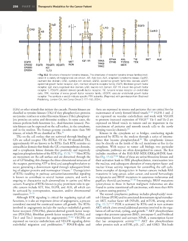
                          • Fig. 15.5  Structure of receptor tyrosine kinases. The structures of receptor tyrosine kinase families impli-
                          cated in a variety of malignancies are shown. AB, Acid box; ALK, anaplastic lymphoma kinase; CadhD,
                          cadherin-like domain;  CRD, cysteine-rich domain;  EGFD, epidermal growth factor-like domain;  EGFR,
                          epidermal growth factor receptor; Eph, member of ephrin receptor family; FGFR, fibroblast growth factor
                          receptor; IgD, immunoglobulin-like domain; LRD, leucine-rich domain; IGF-1R, insulin like growth factor
                          receptor 1; PDGFR, platelet-derived growth factor receptor; TIE, tyrosine kinase receptor on endothelial
                          cells; TRK, member of nerve growth factor receptor family; VEGFR, vascular endothelial growth factor
                          receptor. The symbols α and β indicate specific RTK subunits. (Reprinted with permission from Blackwell
                          Publishing, London CA, Vet Comp Oncol 2:177–193, 2004.)


         (GFs) or other stimuli that initiate the cascade. Protein kinases are   -beta are expressed in stroma and pericytes that are critical for the
         classified as tyrosine kinases (TKs) if they phosphorylate proteins   maintenance of newly formed blood vessels. 81,82  FGFR-1 and -2
         on tyrosine residues or serine/threonine kinases if they phosphory-  are expressed on vascular endothelium and work with VEGFR
                                                                                                 81
         late proteins on serine and threonine residues. In some cases, the   to promote increased expression of VEGF.  Tie-1 and Tie-2 are
         kinases perform both functions (i.e., dual-function kinases). Pro-  expressed on blood vessels in tumors and are important in the
         tein kinases can be expressed on the cell surface, in the cytoplasm,   recruitment of pericytes and smooth muscle cells to the newly
         and in the nucleus. The human genome encodes more than 500   forming vascular channels. 83
         kinases, of which 90 are classified as TKs. 70           Kinases in the cytoplasm act as bridges, conducting signals
            TKs on the cell surface that are activated through binding of   generated by RTKs to the nucleus through a series of interme-
         GFs are called receptor TKs (RTKs). Of the 90 identified TKs,   diates that become phosphorylated.  The cytoplasmic kinases
                                                                                            84
         approximately 60 are known to be RTKs. Each RTK contains an   may be directly on the inside of the cell membrane or free in the
         extracellular domain that binds the GF, a transmembrane domain,   cytoplasm.  With respect to tumor cell biology, two particular
         and a cytoplasmic kinase domain that positively and negatively   cytoplasmic pathways are often dysregulated in cancer. The first
         regulates phosphorylation of the RTK (Fig. 15.5). 71–73  Most RTKs   includes members of the RAS-RAF-MEK-ERK/p38/JNK fami-
         are monomers on the cell surface and are dimerized through the   lies (Fig. 15.6). 85,86  Most of these are serine/threonine kinases and
         act of GF binding; this changes the three-dimensional structure of   their activation leads to ERK phosphorylation, translocation into
         the receptor, permitting ATP to bind and autophosphorylation to   the nucleus, and subsequent alteration of transcription factor and
         occur, generating a downstream signal through subsequent bind-  nuclear kinase activity important for controlling the cell cycle.
         ing of adaptor proteins and nonreceptor kinases.  Dysregulation   Some examples of dysregulation in human cancers include RAS
                                               71
         of RTKs resulting in pathway activation/uncontrolled signaling   mutations in lung cancer, colon cancer, and several hematologic
         is known to contribute to several human cancers, and work is   malignancies and BRAF mutations in cutaneous melanomas and
         ongoing to characterize such abnormalities in canine and feline   papillary thyroid carcinomas. 87–89  Interestingly, BRAF mutations
         cancers. Examples of RTKs known to play prominent roles in spe-  synonymous to those in human malignant melanomas are also
         cific cancers include KIT, Met, EGFR, and ALK, all which can   found in canine transitional cell carcinomas, with more than 80%
         be activated by overexpression, mutation, and/or chromosomal   of tumors testing positive. 90
         translocation. 74–78                                     The second cytoplasmic pathway includes phosphatidyl inosi-
            Although RTK signaling is critical for regulating typical cell   tol-3 kinase (PI3K) and its associated downstream signal transduc-
         functions, it is also an important driver of angiogenesis, a process   ers AKT, nuclear factor κB (NFκB), and mTOR, among others
         considered essential for continued tumor cell growth. The RTKs   (Fig. 15.7). 91,92  PI3K is activated by RTKs and in turn activates
         involved in angiogenesis include the vascluar endothelial growth   AKT, which alters several additional proteins involved in the regu-
         factor receptors (VEGFRs), platelet-derived growth factor recep-  lation of cell survival, cycling, and growth.  AKT phosphorylates
                                                                                                93
         tors (PDGFRs), fibroblast growth factor receptors (FGFRs), and   targets that promote apoptosis (BAD, procaspase-9, and Forkhead
         Tie-1 and Tie-2 (receptors for angiopoietin). 79–82  VEGFRs  are   transcription factors) and activates NFκB, a transcription factor
         expressed on vascular endothelium and VEGFR signaling drives   that has antiapoptotic activity. 91–93  AKT also phosphorylates
         endothelial migration and proliferation.  PDGFR-alpha and   other proteins such as mTOR, p21, p27, and GSK3. This leads
                                          79

