Page 280 - Withrow and MacEwen's Small Animal Clinical Oncology, 6th Edition
P. 280
CHAPTER 15  Molecular/Targeted Therapy of Cancer  259


             Growth
              factor
  VetBooks.ir  Receptor


                   P       P             SOS     RAS   GDP       RAS   GTP
                   P       P  SHC  GRB2

                                                                       RAF  P
                                                                     P
                                                                                  P
                                                                             MEK

                                                                           P               P
                                                                                      ERK
                                                                                   P


                                                                               p90RSK          ELK1

                                                                Nucleus

                                                                                     Gene transcription
                                                                                    cell cycle progression

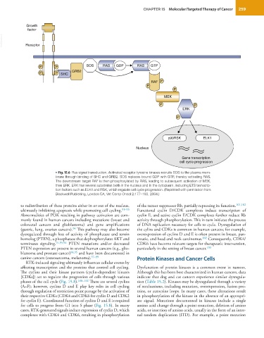
                           • Fig. 15.6  Ras signal transduction. Activated receptor tyrosine kinases recruits SOS to the plasma mem-
                           brane through binding of SHC and GRB2. SOS replaces bound GDP with GTP, thereby activating RAS.
                           The downstream target RAF is then phosphorylated by RAS, leading to subsequent activation of MEK,
                           then ERK. ERK has several substrates both in the nucleus and in the cytoplasm, including ETS transcrip-
                           tion factors such as ELK1 and RSK, which regulate cell cycle progression. (Reprinted with permission from
                           Blackwell Publishing, London CA, Vet Comp Oncol 2:177–193, 2004.)

           to redistribution of these proteins either in or out of the nucleus,   of the tumor suppressor Rb, partially repressing its function. 101,102
           ultimately inhibiting apoptosis while promoting cell cycling. 91–93    Functional cyclin D/CDK complexes induce transcription of
           Abnormalities of PI3K resulting in pathway activation are com-  cyclin E, and active cyclin E/CDK complexes further reduce Rb
           monly found in human cancers including mutations (breast and   activity through phosphorylation. This in turn initiates the process
           colorectal cancers and glioblastoma) and gene amplifications   of DNA replication necessary for cells to cycle. Dysregulation of
                                   94
           (gastric, lung, ovarian cancers).  This pathway may also become   the cylins and CDKs is common in human cancers; for example,
           dysregulated through loss of activity of phosphatase and tensin   overexpression of cyclins D and E is often present in breast, pan-
           homolog (PTEN), a phosphatase that dephosphorylates AKT and   creatic, and head and neck carcinomas. 102  Consequently, CDK4/
           terminates signaling. 91,95,96  PTEN mutations and/or decreased   CDK6 have become relevant targets for therapeutic intervention,
           PTEN expression are present in several human cancers (e.g., glio-  particularly in the setting of breast cancer. 103
           blastoma and prostate cancer) 94,95  and have been documented in
           canine cancers (osteosarcoma, melanoma). 97–99        Protein Kinases and Cancer Cells
             RTK-induced signaling ultimately influences cellular events by
           affecting transcription and the proteins that control cell cycling.   Dysfunction of protein kinases is a common event in tumors.
           The cyclins and their kinase partners (cyclin-dependent kinases   Although this has been best characterized in human cancers, data
           [CDKs]) act to regulate the progression of cells through various   indicate that dog and cat cancers experience similar dysregula-
           phases of the cell cycle (Fig. 15.8). 100–102  There are several cyclins   tion (Table 15.2). Kinases may be dysregulated through a variety
           (A–F); however, cyclins D and E play key roles in cell cycling   of mechanisms, including mutation, overexpression, fusion pro-
           through regulation of restriction point passage by the activation of   teins, or autocrine loops. In many cases, these alterations result
           their respective CDKs (CDK4 and CDK6 for cyclin D and CDK2   in phosphorylation of the kinase in the absence of an appropri-
           for cyclin E). Coordinated function of cyclins D and E is required   ate  signal. Mutations  documented  in  kinases  include  a  single
           for cells to progress from G1 into S phase (Fig. 15.8). In many   amino acid change through a point mutation, deletion of amino
           cases, RTK-generated signals induce expression of cyclin D, which   acids, or insertion of amino acids, usually in the form of an inter-
           complexes with CDK4 and CDK6, resulting in phosphorylation   nal tandem duplication (ITD). For example, a point mutation

