
Page 408 - Withrow and MacEwen's Small Animal Clinical Oncology, 6th Edition
P. 408
386 PART IV Specific Malignancies in the Small Animal Patient
TABLE 21.4 Survival Times of Dogs with Surgically
Treated Mast Cell Tumors According to
a
VetBooks.ir Investigator Histologic Grade Surgery Time (weeks)
Number Percent Months Post Median Survival
of Dogs Alive
Bostock 59
Low grade 39 79 7 NR
Intermediate 30 37 7
grade
High grade 45 15 7
Patnaik 14
Low grade 30 83 48 NR
Intermediate 36 44 48
grade
High grade 17 6 48
Bostock 104
Low grade 19 90 NR >40 b
Intermediate 16 75 >36
grade
High grade 15 27 13
Murphy 84
Low grade 87 100 12 >80 b
Intermediate 199 92 12 >80
grade
High grade 54 46 12 40
Simoes 85
Low grade 33 91 20 NR
Intermediate 35 71 20
grade
High grade 19 42 20
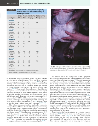
• Fig. 21.3 Subungual undifferentiated mast cell tumor in an English bulldog.
NR, Not reported. As with some mast cell tumors in this location, early lymph node metastasis
a Unclear in these studies if death was due to metastasis or local recurrence. has occurred. (Courtesy D. Vail, University of Wisconsin–Madison.)
b Medians not reached at the time of last follow-up (i.e., >50% alive).
The potential role of KIT dysregulation in MCT prognosis
of argyrophilic nucleolar organizer regions (AgNOR), another was investigated by assessing KIT immunohistochemical staining
surrogate marker of proliferation. These have been correlated patterns on histopathologic specimens. 108 Three distinct patterns
with histologic grade and postsurgical outcome. 79,98 Finally, pro- were identified: membrane, focal/stippled, and diffuse cytoplas-
liferating cell nuclear antigen (PCNA), another indicator of cell mic staining. Although there was some evidence that dogs with
proliferation, has been used to determine the biologic behavior diffuse cytoplasmic KIT staining patterns did not live as long as
of MCTs, although this is probably not as reliable as the other those with other patterns, no group reached an MST and most
markers. 79,98,99 The previously discussed markers of proliferation dogs in each of the KIT staining groups evaluated experienced
all require the use of special stains. In contrast, mitotic index (MI, extremely long postoperative STs. 108 The presence of c-kit activat-
number of mitoses per 10 HPF) in hematoxylin and eosin–stained ing mutations has been associated with a higher rate of local recur-
sections has been used to assess the biologic behavior of canine rence, metastasis, and death from disease, suggesting that KIT
MCTs. In one study, those dogs with tumors possessing an MI <5 dysregulation confers a more aggressive phenotype to MCT. 43,48,49
had a median ST (MST) of 80 months compared with 3 months Finally, investigators have attempted to correlate histologic grad-
for those possessing a MI >5, suggesting that MI is a strong pre- ing of MCT with a combined Ki67/PCNA/AgNOR/KIT immu-
dictor of overall survival for dogs with MCTs. 100 Additional stud- nohistochemical scoring. 109 No significant correlation was found
ies have also found a role for MI in MCT prognosis. 87,97,101–103 for KIT staining and MCT grade, but high Ki67/PCNA/AgNOR
Other cellular assessments have been employed to evaluate the scores all positively correlated with tumor grade (i.e., higher scores
biologic behavior of MCTs. A study of DNA ploidy determined by for higher grade). This suggests that proliferation indices increase
flow cytometric analysis suggested a trend toward shorter survival with increasing grade and are ultimately reflected in the eventual
and higher clinical stage of disease in aneuploid tumors compared biologic behavior of the tumor.
with diploid tumors. 104 Complementary to this, increases in DNA Tumor location has been investigated as a potential prognos-
CNV also appear to be associated with higher grade and shorter tic indicator. 58,110–114 Tumors in the preputial/inguinal area, sub-
postsurgical ST. 51,52 Studies have found a correlation between ungual (nail bed) region (Fig. 21.3), and other mucocutaneous
intratumor microvessel density and invasiveness, MI, and progno- sites, including the oral cavity and perineum, historically have
sis, 89,105 and a correlation between nuclear characteristics (assessed been associated with aggressive behavior. Two reports did not
by computerized morphometry) and outcome and grade. 106,107 show a poorer prognosis for tumors occurring in the inguinal