
Page 409 - Withrow and MacEwen's Small Animal Clinical Oncology, 6th Edition
P. 409
CHAPTER 21 Mast Cell Tumors 387
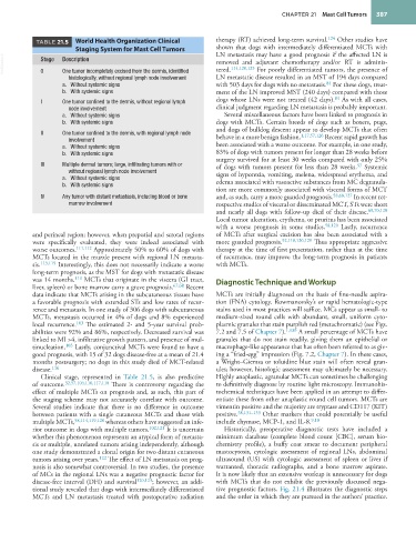
TABLE 21.5 World Health Organization Clinical therapy (RT) achieved long-term survival. 124 Other studies have
Staging System for Mast Cell Tumors shown that dogs with intermediately differentiated MCTs with
LN metastasis may have a good prognosis if the affected LN is
VetBooks.ir Stage Description removed and adjuvant chemotherapy and/or RT is adminis-
For poorly differentiated tumors, the presence of
tered.
114,120,125
0
One tumor incompletely excised from the dermis, identified
histologically, without regional lymph node involvement LN metastatic disease resulted in an MST of 194 days compared
84
a. Without systemic signs with 503 days for dogs with no metastasis. For these dogs, treat-
b. With systemic signs ment of the LN improved MST (240 days) compared with those
84
I One tumor confined to the dermis, without regional lymph dogs whose LNs were not treated (42 days). As with all cases,
node involvement clinical judgment regarding LN metastasis is probably important.
a. Without systemic signs Several miscellaneous factors have been linked to prognosis in
b. With systemic signs dogs with MCTs. Certain breeds of dogs such as boxers, pugs,
and dogs of bulldog descent appear to develop MCTs that often
II One tumor confined to the dermis, with regional lymph node behave in a more benign fashion. 1,17,57,126 Recent rapid growth has
involvement
a. Without systemic signs been associated with a worse outcome. For example, in one study,
b. With systemic signs 83% of dogs with tumors present for longer than 28 weeks before
surgery survived for at least 30 weeks compared with only 25%
III Multiple dermal tumors; large, infiltrating tumors with or of dogs with tumors present for less than 28 weeks. Systemic
57
without regional lymph node involvement signs of hyporexia, vomiting, melena, widespread erythema, and
a. Without systemic signs edema associated with vasoactive substances from MC degranula-
b. With systemic signs
tion are more commonly associated with visceral forms of MCT
IV Any tumor with distant metastasis, including blood or bone and, as such, carry a more guarded prognosis. 58,68,127 In recent ret-
marrow involvement rospective studies of visceral or disseminated MCT, STs were short
and nearly all dogs with follow-up died of their disease. 69,70,128
Local tumor ulceration, erythema, or pruritus has been associated
with a worse prognosis in some studies. 58,120 Lastly, recurrence
and perineal region; however, when preputial and scrotal regions of MCTs after surgical excision has also been associated with a
were specifically evaluated, they were indeed associated with more guarded prognosis. 92,118,120,129 Thus appropriate aggressive
worse outcomes. 111,112 Approximately 50% to 60% of dogs with therapy at the time of first presentation, rather than at the time
MCTs located in the muzzle present with regional LN metasta- of recurrence, may improve the long-term prognosis in patients
sis. 113,115 Interestingly, this does not necessarily indicate a worse with MCTs.
long-term prognosis, as the MST for dogs with metastatic disease
was 14 months. 113 MCTs that originate in the viscera (GI tract, Diagnostic Technique and Workup
liver, spleen) or bone marrow carry a grave prognosis. 67,68 Recent
data indicate that MCTs arising in the subcutaneous tissues have MCTs are initially diagnosed on the basis of fine-needle aspira-
a favorable prognosis with extended STs and low rates of recur- tion (FNA) cytology. Rowmanovsky’s or rapid hematologic-type
rence and metastasis. In one study of 306 dogs with subcutaneous stains used in most practices will suffice. MCs appear as small- to
MCTs, metastasis occurred in 4% of dogs and 8% experienced medium-sized round cells with abundant, small, uniform cyto-
local recurrence. 103 The estimated 2- and 5-year survival prob- plasmic granules that stain purplish red (metachromatic) (see Figs.
abilities were 92% and 86%, respectively. Decreased survival was 7.2 and 7.5 of Chapter 7). 1,130 A small percentage of MCTs have
linked to MI >4, infiltrative growth pattern, and presence of mul- granules that do not stain readily, giving them an epithelial or
tinucleation. 103 Lastly, conjunctival MCTs were found to have a macrophage-like appearance that has often been referred to as giv-
good prognosis, with 15 of 32 dogs disease-free at a mean of 21.4 ing a “fried-egg” impression (Fig. 7.2, Chapter 7). In these cases,
months postsurgery; no dogs in this study died of MCT-related a Wright–Giemsa or toluidine blue stain will often reveal gran-
disease. 116 ules; however, histologic assessment may ultimately be necessary.
Clinical stage, represented in Table 21.5, is also predictive Highly anaplastic, agranular MCTs can sometimes be challenging
of outcome. 32,57,104,110,117,118 There is controversy regarding the to definitively diagnose by routine light microscopy. Immunohis-
effect of multiple MCTs on prognosis and, as such, this part of tochemical techniques have been applied in an attempt to differ-
the staging scheme may not accurately correlate with outcome. entiate these from other anaplastic round cell tumors. MCTs are
Several studies indicate that there is no difference in outcome vimentin positive and the majority are tryptase and CD117 (KIT)
between patients with a single cutaneous MCTs and those with positive. 38,131–133 Other markers that could potentially be useful
multiple MCTs, 58,114,119,120 whereas others have suggested an infe- include chymase, MCP-1, and IL-8. 9,10
rior outcome in dogs with multiple tumors. 102,121 It is uncertain Historically, preoperative diagnostic tests have included a
whether this phenomenon represents an atypical form of metasta- minimum database (complete blood count [CBC], serum bio-
sis or multiple, unrelated tumors arising independently, although chemistry profile), a buffy coat smear to document peripheral
one study demonstrated a clonal origin for two distant cutaneous mastocytosis, cytologic assessment of regional LNs, abdominal
tumors arising over years. 122 The effect of LN metastasis on prog- ultrasound (US) with cytologic assessment of spleen or liver if
nosis is also somewhat controversial. In two studies, the presence warranted, thoracic radiographs, and a bone marrow aspirate.
of MCs in the regional LNs was a negative prognostic factor for It is now likely that an extensive workup is unnecessary for dogs
disease-free interval (DFI) and survival 120,123 ; however, an addi- with MCTs that do not exhibit the previously discussed nega-
tional study revealed that dogs with intermediately differentiated tive prognostic factors. Fig. 21.4 illustrates the diagnostic steps
MCTs and LN metastasis treated with postoperative radiation and the order in which they are pursued in the authors’ practice.