
Page 410 - Withrow and MacEwen's Small Animal Clinical Oncology, 6th Edition
P. 410
388 PART IV Specific Malignancies in the Small Animal Patient
Anatomic site amenable to wide surgical excision?
VetBooks.ir Yes No Expand diagnostics prior to definitive therapy:
1. Biopsy for histologic grade (+/– KIT analysis)
2. Lymph node aspirate
3. Abdominal ultrasound +/– spleen/liver aspirate
4. CBC, biochemistry
Negative prognostic factors
present? (Table 21.1) Yes
If poorly differentiated, highly
No
proliferative or
surgical margins incomplete
Excise with wide surgical margins.
Submit for grade and margin
Complete margins, intermediate
or low grade, and no negative
prognostic indicators
Routine follow-up:
• 1, 3, 6, 9, 12, 15, 18 months
• q 6 months thereafter
• Physical exam and lymph node exam
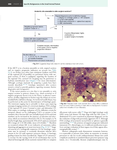
• Fig. 21.4 Suggested diagnostic steps for canine cutaneous mast cell tumors.
If the MCT is in a location amenable to wide surgical excision
and no negative prognostic indicators are present (see Table
21.1), no further tests other than minimum database and FNA
of the regional LN (if possible) are performed before wide sur-
gical excision. If there is ambiguity regarding the location of
the regional LNs, sentinel LN mapping may be performed to
facilitate identification (see Chapter 9 for more information).
Although cytologic methods for assigning a grade have been
recently described, 134–136 histologic assessment after excision
remains critical to provide guidance regarding necessary further
diagnostics and therapeutics.
If the tumor presents at a site that is not amenable to wide
surgical excision or primary closure (e.g., distal extremity) or if
negative prognostic factors exist in the history or physical exami-
nation, ancillary diagnostics to further stage the disease are recom-
mended before definitive therapy. An incisional/needle biopsy may
be performed at this point for determination of histologic grade.
The minimum staging that is advisable in those cases requiring • Fig. 21.5 Regional lymph node aspirate from a dog with a cutaneous
presurgical staging consists of a minimum database, FNA cytology mast cell tumor. Note clustering of mast cells in a background of lympho-
of the regional LN (even if normal in size), and abdominal US. cytes more indicative of true metastasis.
With respect to cytologic evaluation of LNs, definitive criteria for
metastatic disease can be challenging if MCs are present in low effacement with tumor cells). 138 Dogs with high histologic node
numbers; this is because MCs are normally found in LNs and their (HN) scores had shorter STs than dogs with low HN scores. 138
numbers can be increased in the presence of infection and ulcer- Abdominal US is now considered an important diagnostic test for
ation, which are sometimes observed in MCTs. For example, in 56 the evaluation of dogs with potentially aggressive MCT. Although
healthy beagle dogs, approximately 24% of LN aspirates contained FNA cytology of structurally normal livers or spleens is generally
MCs (range of 1–16 MCs/slide, mean of 6.4/slide). 137 Therefore unrewarding, 139,140 the presence of negative prognostic indicators
an occasional solitary MC is not indicative of metastasis; rather, (metastatic LN, clinical signs, etc.) is sufficient justification to per-
clustering and aggregates are more worrisome (Fig. 21.5). 117 Surgi- form cytologic evaluation of these organs even if they appear ultra-
cal removal of a cytologically suspicious LN for histologic assess- sonographically normal. 141
ment may be necessary to accurately determine whether MCs Thoracic radiographs rarely demonstrate metastasis; however,
present in the LN truly represent metastatic disease. A histologic it is reasonable to procure them before an expensive or invasive
grading scheme has been described for “degree of LN metastasis” procedure to rule out occult cardiopulmonary disease that could
to account for the varying levels of LN involvement that can be complicate anesthesia or unrelated disease processes (primary lung
observed histologically (from scattered, isolated MCs to complete tumor, etc.). Occasionally, thoracic lymphadenomegaly may be