
Page 416 - Withrow and MacEwen's Small Animal Clinical Oncology, 6th Edition
P. 416
394 PART IV Specific Malignancies in the Small Animal Patient
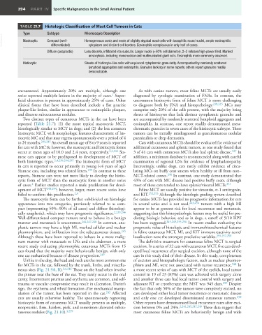
TABLE 21.7 Histologic Classification of Mast Cell Tumors in Cats
Type Subtype Microscopic Description
VetBooks.ir Mastocytic Compact (well- Homogeneous cords and nests of slightly atypical mast cells with basophilic round nuclei, ample eosinophilic
cytoplasm and distinct cell borders. Eosinophils conspicuous in only half of cases.
differentiated)
Diffuse (anaplastic) Less discrete, infiltrated into subcutis. Larger nuclei (>50% cell diameter), 2–3 mitoses/high-power field. Marked
anisocytosis, including mononuclear and multinucleated giant cells. Eosinophils more commonly observed.
Histiocytic Sheets of histiocyte-like cells with equivocal cytoplasmic granularity. Accompanied by randomly scattered
lymphoid aggregates and eosinophils. Granules lacking in some reports, others report granules readily
demonstrable.
encountered. Approximately 20% are multiple, although one As with canine tumors, most feline MCTs are usually easily
1
series reported multiple lesions in the majority of cases. Super- diagnosed by cytologic examination of FNAs. In contrast, the
ficial ulceration is present in approximately 25% of cases. Other uncommon histiocytic form of feline MCT is more challenging
clinical forms that have been described include a flat pruritic to diagnose both by FNA and histopathology. 239,245 MCs may
plaque-like lesion, similar in appearance to eosinophilic plaques, comprise only 20% of the cells present, with the majority being
and discrete subcutaneous nodules. sheets of histiocytes that lack distinct cytoplasmic granules and
Two distinct types of cutaneous MCTs in the cat have been are accompanied by randomly scattered lymphoid aggregates and
reported (Table 21.7): (1) the more typical mastocytic MCT, eosinophils. In contrast, one report readily demonstrated meta-
histologically similar to MCT in dogs; and (2) the less common chromatic granules in seven cases of the histiocytic subtype. These
histiocytic MCT, with morphologic features characteristic of his- tumors can be initially misdiagnosed as granulomatous nodular
tiocytic MC and that may regress spontaneously over a period of 4 panniculitis or deep dermatitis.
to 24 months. 239,245 An overall mean age of 8 to 9 years is reported Cats with cutaneous MCTs should be evaluated for evidence of
for cats with MCTs; however, the mastocytic and histiocytic forms additional cutaneous and splenic tumors, as one study found that
occur at mean ages of 10.0 and 2.4 years, respectively. 2,4,244 Sia- 3 of 41 cats with cutaneous MCTs also had splenic disease. 247 In
mese cats appear to be predisposed to development of MCT of addition, a minimum database is recommended along with careful
both histologic types. 2,4,239,244,245 The histiocytic form of MCT examination of regional LNs for evidence of lymphadenopathy.
in cats is reported to occur primarily in young (<4 years of age) Interestingly, unlike dogs, cats rarely exhibit evidence of circu-
Siamese cats, including two related litters. 239 In contrast to these lating MCs on buffy coat smears when healthy or ill from non–
reports, Siamese cats were not more likely to develop the histio- MCT-related causes. 250 In contrast, one study demonstrated that
cytic form of MCT than the mastocytic form in another series 43% of cats with MC disease had positive buffy coats, although
4
of cases. Earlier studies reported a male predilection for devel- most of these cats tended to have splenic/visceral MCTs. 251
opment of MCT 243,244 ; however, larger, more recent series have Feline MCT are usually positive for vimentin, α-1 antitrypsin,
failed to confirm this predilection. 2,4 and KIT. 252,253 Although the histologic grading system described
The mastocytic form can be further subdivided on histologic for canine MCTs has provided no prognostic information for cats
appearance into two categories, previously referred to as com- in several series and is not used, 243,254 tumors with a high MI
pact (representing 50%–90% of all cases) and diffuse (histologi- appear to be at greatest risk for local recurrence and metastasis,
cally anaplastic), which may have prognostic significance. 2,239,246 suggesting that this histopathologic feature may be useful for pre-
Well-differentiated compact tumors tend to behave in a benign dicting biologic behavior, and as in dogs, a cutoff of 5/10 HPF
manner and metastasis is uncommon. 244,247,248 In contrast, ana- has been suggested. 245,249,255,256 In recent studies evaluating the
plastic tumors may have a high MI, marked cellular and nuclear prognostic value of histologic and immunohistochemical features
pleomorphism, and infiltration into the subcutaneous tissues. 245 in feline cutaneous MCT, MI, and KIT immunoreactivity score/
Although these have been reported to behave in a more malig- localization were the strongest predictive variables. 255,257,258
nant manner with metastasis to LNs and the abdomen, a more The definitive treatment for cutaneous feline MCT is surgical
recent study evaluating pleomorphic cutaneous MCTs from 15 excision. In a series of 32 cats with cutaneous MCT, five cats devel-
cats found that the majority were behaviorally benign, with only oped local recurrence after surgical excision, although none of the
one cat euthanized because of disease progression. 249 cats in this study died of their disease. In this study, completeness
Unlike in the dog, the head and neck are the most common site of excision and histopathologic factors, such as nuclear pleomor-
for MCTs in the cat, followed by the trunk, limbs, and miscella- phism and MI, were not associated with tumor recurrence. 248 In
neous sites (Fig. 21.9A, B). 2,4,244 Those on the head often involve a more recent series of cats with MCT of the eyelids, local tumor
the pinnae near the base of the ear. They rarely occur in the oral control in 19 of 23 (83%) cats was achieved with surgery alone
cavity. Intermittent pruritus and erythema are common, and self- and another three cats had local tumor control with surgery and
trauma or vascular compromise may result in ulceration. Darier’s adjuvant RT or cryotherapy; the MST was 945 days. 259 Despite
sign, the erythema and wheal formation after mechanical manip- the fact that only 50% of the tumors were completely excised, no
ulation of the tumor, has been reported in the cat. 243 Affected cats developed either local tumor recurrence or metastatic disease
cats are usually otherwise healthy. The spontaneously regressing and only one cat developed disseminated cutaneous tumors. 259
histiocytic form of cutaneous MCT usually presents as multiple, Other reports have demonstrated local recurrence rates after exci-
nonpruritic, firm, hairless, pink, and sometimes ulcerated subcu- sion between 0% and 24%. 2,244,245,247,249 These data suggest that
taneous nodules (Fig. 21.10). 4,239 most cutaneous feline MCTs are behaviorally benign and wide