
Page 418 - Withrow and MacEwen's Small Animal Clinical Oncology, 6th Edition
P. 418
396 PART IV Specific Malignancies in the Small Animal Patient
VetBooks.ir
A B
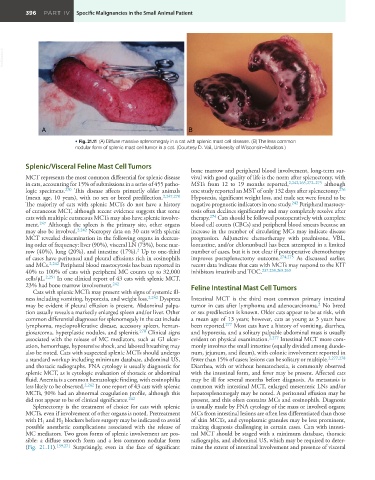
• Fig. 21.11 (A) Diffuse massive splenomegaly in a cat with splenic mast cell disease. (B) The less common
nodular form of splenic mast cell tumor in a cat. (Courtesy D. Vail, University of Wisconsin–Madison.)
Splenic/Visceral Feline Mast Cell Tumors
bone marrow and peripheral blood involvement, long-term sur-
MCT represents the most common differential for splenic disease vival with good quality of life is the norm after splenectomy, with
in cats, accounting for 15% of submissions in a series of 455 patho- MSTs from 12 to 19 months reported, 2,242,265,272–275 although
logic specimens. 270 This disease affects primarily older animals one study reported an MST of only 132 days after splenectomy. 276
(mean age, 10 years), with no sex or breed predilection. 2,247,270 Hyporexia, significant weight loss, and male sex were found to be
The majority of cats with splenic MCTs do not have a history negative prognostic indicators in one study. 242 Peripheral mastocy-
of cutaneous MCT, although recent evidence suggests that some tosis often declines significantly and may completely resolve after
cats with multiple cutaneous MCTs may also have splenic involve- therapy. 274 Cats should be followed postoperatively with complete
ment. 247 Although the spleen is the primary site, other organs blood cell counts (CBCs) and peripheral blood smears because an
may also be involved. 2,242 Necropsy data on 30 cats with splenic increase in the number of circulating MCs may indicate disease
MCT revealed dissemination in the following organs in decreas- progression. Adjunctive chemotherapy with prednisone, VBL,
ing order of frequency: liver (90%), visceral LN (73%), bone mar- lomustine, and/or chlorambucil has been attempted in a limited
2
row (40%), lung (20%), and intestine (17%). Up to one-third number of cases, but it is not clear if postoperative chemotherapy
of cases have peritoneal and pleural effusions rich in eosinophils improves postsplenectomy outcome. 274,275 As discussed earlier,
and MCs. 2,242 Peripheral blood mastocytosis has been reported in recent data indicate that cats with MCTs may respond to the KIT
40% to 100% of cats with peripheral MC counts up to 32,000 inhibitors imatinib and TOC. 237,238,268,269
cells/μL. 2,251 In one clinical report of 43 cats with splenic MCT,
23% had bone marrow involvement. 242 Feline Intestinal Mast Cell Tumors
Cats with splenic MCTs may present with signs of systemic ill-
ness including vomiting, hyporexia, and weight loss. 2,242 Dyspnea Intestinal MCT is the third most common primary intestinal
may be evident if pleural effusion is present. Abdominal palpa- tumor in cats after lymphoma and adenocarcinoma. No breed
2
tion usually reveals a markedly enlarged spleen and/or liver. Other or sex predilection is known. Older cats appear to be at risk, with
common differential diagnoses for splenomegaly in the cat include a mean age of 13 years; however, cats as young as 3 years have
lymphoma, myeloproliferative disease, accessory spleen, heman- been reported. 277 Most cats have a history of vomiting, diarrhea,
giosarcoma, hyperplastic nodules, and splenitis. 270 Clinical signs and hyporexia, and a solitary palpable abdominal mass is usually
associated with the release of MC mediators, such as GI ulcer- evident on physical examination. 2,277 Intestinal MCT more com-
ation, hemorrhage, hypotensive shock, and labored breathing may monly involves the small intestine (equally divided among duode-
also be noted. Cats with suspected splenic MCTs should undergo num, jejunum, and ileum), with colonic involvement reported in
a standard workup including minimum database, abdominal US, fewer than 15% of cases; lesions can be solitary or multiple. 2,277,278
and thoracic radiographs. FNA cytology is usually diagnostic for Diarrhea, with or without hematochezia, is commonly observed
splenic MCT, as is cytologic evaluation of thoracic or abdominal with the intestinal form, and fever may be present. Affected cats
fluid. Anemia is a common hematologic finding, with eosinophilia may be ill for several months before diagnosis. As metastasis is
less likely to be observed. 2,242 In one report of 43 cats with splenic common with intestinal MCT, enlarged mesenteric LNs and/or
MCTs, 90% had an abnormal coagulation profile, although this hepatosplenomegaly may be noted. A peritoneal effusion may be
did not appear to be of clinical significance. 242 present, and this often contains MCs and eosinophils. Diagnosis
Splenectomy is the treatment of choice for cats with splenic is usually made by FNA cytology of the mass or involved organs;
MCTs, even if involvement of other organs is noted. Pretreatment MCs from intestinal lesions are often less differentiated than those
with H and H blockers before surgery may be indicated to avoid of skin MCTs, and cytoplasmic granules may be less prominent,
2
1
possible anesthetic complications associated with the release of making diagnosis challenging in certain cases. Cats with intesti-
MC mediators. Two gross forms of splenic involvement are pos- nal MCT should be staged with a minimum database, thoracic
sible: a diffuse smooth form and a less common nodular form radiographs, and abdominal US, which may be required to deter-
(Fig. 21.11). 139,271 Surprisingly, even in the face of significant mine the extent of intestinal involvement and presence of visceral