
Page 518 - Withrow and MacEwen's Small Animal Clinical Oncology, 6th Edition
P. 518
496 PART IV Specific Malignancies in the Small Animal Patient
VetBooks.ir
A B
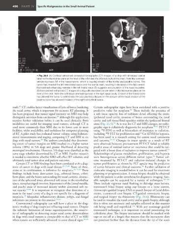
• Fig. 24.4 (A) Contrast-enhanced computed tomography (CT) images of a dog with sinonasal cancer
taken in the transverse plane at the level of the orbit and the olfactory bulb of the brain. Note the contrast-
enhancing mass (M) in the nasopharynx, which is causing erosion of the frontal and palatine bones. The
tumor has invaded the left retroorbital space and the cranial vault, resulting in deviation of the falx cerebri.
Noncontrast-enhancing material in the left frontal sinus (S) suggests accumulation of the nasal exudates.
(B) Noncontrast-enhanced CT images of a dog with sinonasal cancer taken in the transverse plane at the
level of the orbit. Note the soft tissue attenuating mass in the right nasal cavity. Erosion of the frontal bone
has allowed the tumor to extend into the subcutaneous tissues on the dorsum of the head; erosion of the
palatine bone has allowed invasion of the right retroorbital space.
85
vault. CT enables better visualization of lysis of bones bordering Certain radiographic signs have been correlated with a positive
92
the nasal cavity, which is important for accurate RT planning. It predictive value for neoplasia. These include the presence of
has been proposed that tumor signal intensity on MRI may help a soft tissue opacity, loss of turbinate detail affecting the entire
86
distinguish sarcomas from carcinomas, although this application ipsilateral nasal cavity, invasion of bones surrounding the nasal
requires further validation before it can be used clinically. Both cavity, and soft tissue/fluid opacities within the ipsilateral frontal
92
modalities are useful for imaging nasal tumors, although CT is sinus (Fig. 24.5). As is true for CT and MRI changes, no radio-
used more commonly than MRI due to its lower cost in most graphic sign is definitively diagnostic for neoplasia. 88,92 PET/CT
18
facilities, wider availability, and usefulness for computer planning using F-FDG as well as biomarkers of resistance to radiation,
18
61
of RT. A pilot study has evaluated tumor volume using bidimen- including F-FLT for proliferation and Cu-ATSM for hypoxia,
sional measurements and staging, comparing CT and MRI in six has been used in a research setting for canine nasal carcinoma
90
dogs with nasal tumors. The authors concluded that determin- and sarcoma. 93–97 Changes in tracer uptake as a result of RT
ing extent of tumor margins on MRI resulted in a higher tumor were observed; however, pretreatment PET/CT failed to reliably
volume (18%) in 5/6 dogs and greater likelihood of detecting predict areas of residual tumor or recurrence that could be tar-
97
meningeal involvement. However, 5/6 dogs were classified as the geted with a boost dose of radiation to improve tumor control.
same stage whether determined by CT or MRI. Further research Relationships of glucose metabolism, proliferation, and hypoxia
94
is needed to determine whether MRI will affect RT volumes, and were heterogeneous across different tumor types. Tumor vol-
ultimately total tumor dose and patient outcome. ume measured by PET-CT and radiation-induced changes in
Certain CT or MRI findings have been correlated with a diag- tumor proliferation as shown by FLT uptake may be predictive
96
nosis of cancer in dogs with nasal disease. 49,75,89 None of these, of tumor behavior and clinical outcome. Further validation of
alone or in combination, is definitive for neoplasia. 49,75,89 These molecular imaging is needed before it can be used for treatment
findings include bony destruction (e.g., ethmoid bones, cribri- planning or prognostication. A tissue biopsy should be obtained
form plate, and the bones surrounding the nasal cavities), destruc- while the patient is under anesthesia for diagnostic imaging. Suit-
tion of the sphenoid sinus, abnormal soft tissue in the retrobulbar able samples can be acquired by a variety of techniques. 98–100
space, nasopharyngeal invasion, hyperostosis of the lateral maxilla, These include vigorous nasal flushing to dislodge mass lesions,
and patchy areas of increased density within abnormal soft tis- transnostril blind biopsy using cup forceps or a bone curette,
sue opacity. 49,89 It is important to recognize that detection of a rhinoscopic-guided biopsy, FNA or punch biopsy of facial defor-
mass in the nasal cavity of a dog is not specific for a diagnosis of mities, transnostril core biopsy, CT-guided stereotactic biopsy,
49
neoplasia. Idiopathic inflammatory disease, polyps, and fungal and surgical biopsy via rhinotomy (Fig. 24.6). Rhinoscopy can
infections can present in this manner. 49,64 be used to visualize the nasal cavity and to guide biopsy, although
Conventional radiography can still have a place in the diag- this is often not necessary and samples collected in this manner
nostic workup of dogs suspected of having nasal tumors. Despite risk being small and superficial. 101 With any nonimage guided
the inherent limitation of tissue superimposition, the sensitiv- transnostril technique, it is important to avoid penetrating the
ity of radiography in detecting major nasal cavity abnormalities cribriform plate. The biopsy instrument should be marked with
in dogs with nasal tumors is comparable to that of CT or MRI tape or cut off at a length that ensures that the instrument does
when tumors are sufficiently advanced to cause clinical signs. 88,91 not penetrate farther than the distance from the tip of the nares