
Page 520 - Withrow and MacEwen's Small Animal Clinical Oncology, 6th Edition
P. 520
498 PART IV Specific Malignancies in the Small Animal Patient
TABLE 24.1 Modified Adams Clinical Staging Method
for Nasosinal Tumors 106
VetBooks.ir Stage 1 Confined to one nasal passage, paranasal sinus or
frontal sinus, with no bone involvement beyond
turbinates
Stage 2 Any bone involvement (beyond turbinates), but with no
evidence of orbit/subcutaneous/submucosal mass
Stage 3 Orbit involved, or nasopharyngeal, or subcutaneous, or
submucosal mass
Stage 4 Tumor-causing lysis of the cribriform plate
number, the typical patient population with nasal tumors is
older, and more thorough screening if treatment is being con-
sidered may be warranted.
Hematologic and biochemical findings generally are noncon-
tributory in dogs with sinonasal tumors. In rare cases, paraneoplas-
tic erythrocytosis and hypercalcemia have been documented. 112–114
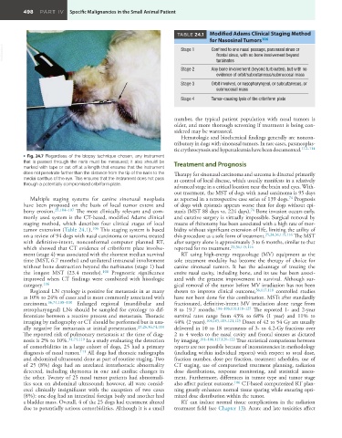
• Fig. 24.7 Regardless of the biopsy technique chosen, any instrument
that is passed through the naris must be measured; it also should be Treatment and Prognosis
marked with tape or cut off at a length that ensures that the instrument
does not penetrate farther than the distance from the tip of the naris to the Therapy for sinonasal carcinoma and sarcoma is directed primarily
medial canthus of the eye. This ensures that the instrument does not pass at control of local disease, which usually manifests in a relatively
through a potentially compromised cribriform plate. advanced stage in a critical location near the brain and eyes. With-
out treatment, the MST of dogs with nasal carcinoma is 95 days
Multiple staging systems for canine sinonasal neoplasia as reported in a retrospective case series of 139 dogs. Prognosis
74
have been proposed on the basis of local tumor extent and of dogs with epistaxis appears worse than for dogs without epi-
bony erosion. 29,104–107 The most clinically relevant and com- staxis (MST 88 days vs. 224 days). Bone invasion occurs early,
74
monly used system is the CT-based, modified Adams clinical and curative surgery is virtually impossible. Surgical removal by
staging method, which describes four clinical stages of local means of rhinotomy has been associated with a high rate of mor-
tumor extension (Table 24.1). 106 This staging system is based bidity without significant extension of life, limiting the utility of
on a review of 94 dogs with nasal carcinoma or sarcoma treated this procedure as a sole form of treatment. 25,34,36,115,116 The MST
with definitive-intent, nonconformal computer planned RT, after surgery alone is approximately 3 to 6 months, similar to that
which showed that CT evidence of cribriform plate involve- reported for no treatment. 25,36,115,116
ment (stage 4) was associated with the shortest median survival RT using high-energy megavoltage (MV) equipment as the
time (MST, 6.7 months) and unilateral intranasal involvement sole treatment modality has become the therapy of choice for
without bone destruction beyond the turbinates (stage 1) had canine sinonasal tumors. It has the advantage of treating the
the longest MST (23.4 months). 106 Prognostic significance entire nasal cavity, including bone, and its use has been associ-
improved when CT findings were combined with histologic ated with the greatest improvement in survival. Although sur-
category. 106 gical removal of the tumor before MV irradiation has not been
Regional LN cytology is positive for metastasis in as many shown to improve clinical outcome, 36,117,118 controlled studies
as 10% to 24% of cases and is most commonly associated with have not been done for this combination. MSTs after standardly
carcinoma. 36,74,108–110 Enlarged regional (mandibular and fractionated, definitive-intent MV irradiation alone range from
retropharyngeal) LNs should be sampled for cytology to dif- 8 to 19.7 months. 104–106,110,118–125 The reported 1- and 2-year
ferentiate between a reactive process and metastasis. Thoracic survival rates range from 43% to 68% (1 year) and 11% to
imaging by radiography or CT should be performed but is usu- 44% (2 years). 104,105,119–121,125 Doses of 42 to 54 Gy are usually
ally negative for metastasis at initial presentation. 25,26,36,74,110 delivered in 10 to 18 treatments of 3- to 4.2-Gy fractions over
The reported risk of pulmonary metastasis at the time of diag- 2 to 4 weeks to the nasal cavity and frontal sinuses as dictated
nosis is 2% to 10%. 34,74,110 In a study evaluating the detection by imaging. 104–106,117,120–122 True statistical comparisons between
of comorbidities in a large cohort of dogs, 25 had a primary reports are not possible because of inconsistencies in methodology
diagnosis of nasal tumor. 111 All dogs had thoracic radiographs (including within individual reports) with respect to total dose,
and abdominal ultrasound done as part of routine staging. Two fraction number, dose per fraction, treatment schedules, use of
of 25 (8%) dogs had an unrelated intrathoracic abnormality CT staging, use of computerized treatment planning, radiation
detected, including thymoma in one and cardiac changes in dose distributions, response monitoring, and statistical assess-
the other. Twenty of 25 nasal tumor patients had abnormali- ment. Furthermore, differences in tumor type and tumor stage
ties seen on abdominal ultrasound; however, all were consid- also affect patient outcome. 106 CT-based computerized RT plan-
ered clinically insignificant with the exception of two cases ning greatly enhances normal tissue sparing while ensuring opti-
(8%): one dog had an intestinal foreign body and another had mized dose distribution within the tumor.
a bladder mass. Overall, 4 of the 25 dogs had treatment altered RT can induce normal tissue complications in the radiation
due to potentially serious comorbidities. Although it is a small treatment field (see Chapter 13). Acute and late toxicities affect