
Page 530 - Withrow and MacEwen's Small Animal Clinical Oncology, 6th Edition
P. 530
508 PART IV Specific Malignancies in the Small Animal Patient
of these cats had a primary pulmonary carcinoma. 290 Nonspecific
signs, such as lethargy and hyporexia, can be seen in 14% to 43% Diagnostics
Vomiting/regurgita-
285–289
and 19% to 71% of cats, respectively.
VetBooks.ir tion and diarrhea are seen in approximately 19% of cats with pri- Clinical Laboratory Findings
A complete blood count and serum chemistry panel are unlikely
285,286
mary pulmonary neoplasia.
Abnormal physical examination findings consistent with pul- to signal the presence of a pulmonary mass. 265,304 These diag-
monary neoplasia are often not seen. 282 Increased bronchovesicu- nostics, however, are essential to the preanesthetic and overall
lar sounds may be auscultated in dogs with extensive pulmonary evaluation of a patient undergoing treatment for a pulmonary
involvement. 282 As pleural effusion can occur in many cases, dull neoplasm. In one study, neutrophilia was noted in 50% of dogs
lung and heart sounds may also be noted. 281,285,286 Metastasis to with HO. 300
the nervous system has been reported in several cases of primary The presence of pleural effusion at diagnosis is less common in
pulmonary tumors and neurologic abnormalities can be diag- dogs than cats. When pleural effusion is noted, a sample should
nosed on physical examination. 291–294 be obtained via thoracocentesis. The pleural fluid tends to be a
Lameness can occur in dogs and cats associated with primary clear or blood-tinged modified transudate. 281,285 Fourteen percent
pulmonary neoplasia for a variety of reasons. HO is a para- to 30% of cats with primary pulmonary tumors have concurrent
neoplastic syndrome commonly associated with primary and pleural effusion. 265,287–289 In one study, 13 of 26 cats underwent
metastatic lung tumors, although other malignant and nonma- thoracocentesis to obtain fluid for analysis; fluid was diagnostic
lignant diseases have resulted in HO. 283,295–300 The disease is for a primary lung tumor in 12 of these cats. 265 However, in a sep-
characterized by periosteal new bone formation at a site distant arate study, fluid analysis was diagnostic for a malignant neoplasm
to the primary tumor. Dogs with HO tend to present with sev- in only one of eight cats. 285 Of three dogs with pleural effusion in
eral clinical abnormalities including limb swelling, lameness, one study, one dog was diagnosed with carcinoma based on evalu-
ocular signs, and lethargy. 300 The prevalence of ocular signs ation of fluid obtained by thoracocentesis. 281
(occurring in 23 of 30 dogs) in a recent study evaluating HO Bronchoalveolar lavage (BAL) and transtracheal washes have
was an interesting finding; an exact association between HO been advocated as a method of diagnosing pulmonary neopla-
and ocular signs was not able to be determined retrospectively sia. 281,305–307 In a series of dogs that underwent BAL to aid in the
in that cohort, but it is important to evaluate for this in dogs diagnosis of respiratory tract diseases, carcinomas were identified
with HO. 300 Lameness may improve with removal of the lung in 14 dogs. Of those 14 cases, the BAL was definitive, supportive,
tumor. 295,298 or not helpful in eight, four, and two dogs, respectively. 306 Trans-
Several reports of cats with concurrent pulmonary neoplasia and tracheal washes have been less successful; of six dogs in one study
digit metastasis can be found in the veterinary literature; this phe- with confirmed primary pulmonary neoplasia, none of the washes
nomenon has been noted with both pulmonary adenocarcinoma and yielded neoplastic cells. 281
SCC. 279,301–304 Cats naturally have significant blood flow to their
digits to allow for heat loss and it has been theorized that this flow Thoracic Radiographs
and the ability of pulmonary tumors to metastasize hematologically
301
may work together to produce this unique metastatic pattern; The majority of pulmonary tumors are diagnosed on thoracic
however, other factors contributing to a favorable metastatic micro- radiographs (Fig. 24.12) and (Fig. 24.13). When evaluating 277
environment are likely to be involved. Of 36 cats with metastatic dogs from two large case series, 83% of pulmonary tumors were
bronchogenic carcinoma to the digit in one study, all presented for visible on thoracic radiographs. 281,283 Radiographic evidence of
lameness and none had respiratory signs. The authors of that study solitary or multiple pulmonary masses are present in 67% to 91%
304
concluded that thoracotomy with lung lobectomy and digital ampu- of cats with primary pulmonary tumors. 265,286
tation should not be recommended as nonrespiratory disease often Several studies have evaluated tumor location and number
progressed and metastatic lesions in other digits resulted in contin- within the lungs. 256,282,283 Single and multiple masses were found
304
ued lameness. The MST for cats undergoing digit amputation was in 54% to 87% and 13% to 37% of dogs, respectively, in two
304
only 67 days. studies. 282,283 A side predilection has not been reported with the
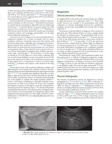
• Fig. 24.12 Right lateral radiograph and ultrasound image of a pulmonary carcinoma (noted by black
arrow) in the left caudal lung lobe of a cat.