
Page 531 - Withrow and MacEwen's Small Animal Clinical Oncology, 6th Edition
P. 531
CHAPTER 24 Tumors of the Respiratory System 509
left and right lung lobes affected in 24% to 50% and 50% to 76% (lobar or segmental consolidations involving one or more
of dogs, respectively. 256,282 Clinical signs were not noted until the lobes), and diffuse (involved most of both lungs). 308 In the
261
focal group, 65% demonstrated solitary masses and 35% dem-
pulmonary tumor grew to at least 3 cm in size in another study.
VetBooks.ir location was determined radiographically in all cases. 265 Tumors onstrated multiple masses. 308 Recently, a study in dogs aimed
In a study of 86 cats with primary pulmonary neoplasia, the
to use radiographic features to distinguish between tumor types
were left-sided in 26 cats, right-sided in 27 cats, and bilateral in 33 and compared pulmonary adenocarcinoma, bronchoalveolar
cats. 265 In 45 cats, the tumors were found in a single lung lobe and carcinoma, and HS. 309 In this study, HSs were significantly
the right and left caudal lung lobes were more commonly involved larger than carcinomas and were more commonly found in the
(34) than the right and left cranial (9) or right middle lung lobes right middle (43%) and left cranial (38%) lung lobes. 309 Addi-
(2). 265 In one study, 19 cats had a single lung lobe affected whereas tionally, 57% of dogs with HS had internal air bronchograms.
two cats had multiple lesions. 286 Of 17 cats in a separate study, all Adenocarcinomas were more often diagnosed in the left caudal
single lesions were left-sided; however, 10 of these cats had mul- lung lobe (29%). 309 Another study noted that in 19 of 29 dogs
tiple lesions (both right and left-sided). 285 with pulmonary HS, the mass was located in the right middle
The radiographic pattern of primary pulmonary neoplasia lung lobe and this was significantly more common than other
has been variably reported in dogs and cats. Three radiographic locations. 310
patterns were described in a study evaluating 41 cats with pri-
mary pulmonary tumors: focal (nodules or masses), localized Thoracic Ultrasound
Thoracic ultrasound may be employed to assess pulmonary neo-
plasia or to obtain a sample of tissue via FNA or pretreatment
biopsy. The ultrasonographic appearance of pulmonary neopla-
sia has been described in several studies. 311–313 Pulmonary masses
may be hypoechoic or exhibit variable echogenicity, and tumors
are generally considered to have both a lack of discernable bronchi
and normal branching vessels. 311,312
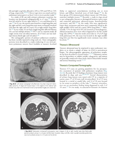
Thoracic Computed Tomography
Thoracic CT scans are gaining popularity for the preopera-
tive assessment of patients with pulmonary neoplasia (Fig.
24.14). Recently the CT findings of primary lung tumors were
described in dogs and cats. 287,288,314 Several characteristics
of solitary lung tumors were noted with CT in a majority of
canine cases; 17 of 18 were well circumscribed, 16 of 19 were
located in a cranial or caudal lobe, and 14 of 18 were located
in the center to periphery of the lobe. 314 Internal mineraliza-
• Fig. 24.13 Left lateral radiograph of a Bernese mountain dog with histio-
cytic sarcoma. A large soft tissue mass in the region of the ventral right tion was an uncommon finding being diagnosed in only 3 of
middle lung lobe and tracheobronchial lymphadenomegaly are observed. 19 cases. 314 In one study, no association between the features
A B
• Fig. 24.14 Transverse computed tomography scan images of (A) a right caudal lung lobe histiocytic
sarcoma (black arrow) in a 9-year-old Bernese mountain dog; and (B) a right caudal lung lobe pulmonary
carcinoma (black arrow) in a 10-year-old fox terrier.