
Page 552 - Withrow and MacEwen's Small Animal Clinical Oncology, 6th Edition
P. 552
530 PART IV Specific Malignancies in the Small Animal Patient
VetBooks.ir
A B
C D
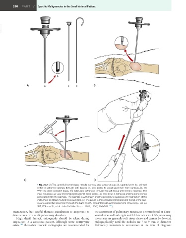
• Fig. 25.2 (A) The Jamshidi bone biopsy needle: cannula and screw-on cap (d), tapered point (b), pointed
stylet to advance cannula through soft tissues (c), and probe to expel specimen from cannula (d). (B)
With the stylet locked in place, the cannula is advanced through the soft tissue until bone is reached. The
inset is a close-up view showing stylet against bone cortex. (C) The stylet is removed and the bone cortex
penetrated with the cannula. The cannula is withdrawn and the procedure repeated with redirection of the
instrument to obtain multiple core samples. (D) The probe is then inserted retrograde into the tip of the can-
nula to expel the specimen through the base (inset). (Reprinted with permission from Powers BE, LaRue
SM, Withrow SJ, et al. J Am Vet Med Assoc. 1988; 193(2):206–207. 140 )
examination, but careful thoracic auscultation is important to the assessment of pulmonary metastasis: a ventrodorsal or dorso-
detect concurrent cardiopulmonary disorders. ventral view and both right and left lateral views. OSA pulmonary
High detail thoracic radiographs should be taken during metastases are generally soft tissue dense and cannot be detected
inspiration in a conscious patient. Although some controversy radiographically until the nodules are 7 to 9 mm in diameter.
exists, 142 three-view thoracic radiographs are recommended for Pulmonary metastasis is uncommon at the time of diagnosis