
Page 553 - Withrow and MacEwen's Small Animal Clinical Oncology, 6th Edition
P. 553
CHAPTER 25 Tumors of the Skeletal System 531
secondary sites considered highly suspect of bony metastasis in
7.8% of (399) cases; however, most suspected lesions were not
confirmed histologically.
150
A recent pilot study compared the
VetBooks.ir utility of survey radiography, whole body CT, and nuclear scintig-
raphy for the detection of OSA metastasis in 15 dogs.
119
Nuclear
scintigraphy was found to be the most useful modality for the
detection of occult bone metastases; however, false positives did
occur, and the diagnosis of metastasis was based on a gold stan-
dard reference constructed using all available imaging modalities,
history, and signalment rather than histopathologic confirmation.
A Abdominal ultrasound has been recommended by some veteri-
nary oncologists to stage for visceral organ metastasis. Two recent
retrospective studies documented abdominal metastases in 0% to
2.5% of cases, with sites of metastasis including the kidney, liver,
and iliac LNs. 153,154 In four of 80 cases (5%), a second primary
neoplasm was diagnosed, and in two of these cases, the mass was
palpable on physical examination. Both studies suggest abdomi-
nal ultrasound is a low-yield diagnostic test but is warranted in
patients with palpable abdominal abnormalities.
Surgical Staging
A surgical staging system for sarcomas of the skeleton has been
C
devised for people. 155 This system is based on the histologic grade
B (G), the anatomic setting of the primary tumor (T), and regional
or distant metastasis (M). There are three stages: stage I—low-
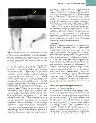
• Fig. 25.3 Scintigraphic view of a distal radial osteosarcoma lesion in a grade (G ) lesions without metastasis; stage II—high-grade (G )
2
1
dog after technetium-99M-hydroxymethylene diphosphonate injection. lesions without metastasis; and stage III—lesions with regional or
(A) Lateral radiograph demonstrating mixed osteolytic/osteoblastic bone distant metastasis regardless of histologic grade. The stages are sub-
lesion affecting distal radius (yellow arrowhead). (B) Anterior posterior and divided by the anatomic setting with A being intracompartmen-
(C) lateral bone scintigraphy images 90 minutes postintravenous injection tal (T ) and B being extracompartmental (T ). According to this
2
1
demonstrate preferential accumulation of technetium-99M at focal site of system, most dogs with OSA present with stage IIB disease. CT
increased bone turnover.
or PET/CT can be used to evaluate the degree of bone involve-
ment from a primary bone tumor and distant metastasis. 145,156
(less than 10% of dogs). Advanced imaging (e.g., CT, MRI, PET/ Scintigraphy has also been used to determine extent of local dis-
CT) may play a role in patient staging and is used to evaluate ease, but in one study, scintigraphy overestimated the length of
for pulmonary metastases and for evaluation of tumor vascularity, OSA disease in LSS patients by 30%. 157 CT may be useful to plan
soft tissue and medullary involvement/tumor size, and response surgical margins, especially for tumors located in the axial skel-
to treatment. 143–145 Multiple studies have demonstrated increased eton; however, one study reported that survey radiographs were as
sensitivity of thoracic CT compared with thoracic radiography accurate as advanced imaging (CT, MRI) in predicting true length
for the detection of pulmonary metastases; 118,119,146,147 however, of tumor involvement. 158 In contrast, it is anticipated that PET/
published treatment and patient outcomes are generally based CT, with its dual molecular and anatomic imaging capabilities,
on patients staged by thoracic radiographs. As advanced imaging will have high sensitivity for assessing local tumor size, as well as
becomes more commonplace for staging dogs with OSA, com- detecting the presence of distant metastases.
parisons to previous protocols will be subject to stage-migration
and lead-time bias due to earlier detection of metastases. Known or Suggested Prognostic Factors
Bone survey radiography may be useful for identifying second
25
skeletal sites of OSA. Bone survey radiographs include lateral Anatomic Location and Signalment
radiographs of all bones in the body and a ventrodorsal projec- In a multiinstitutional study of 162 dogs with appendicular OSA
tion of the pelvis using standard radiographic technique appro- treated with amputation alone, dogs younger than 5 years of age
priate for the region radiographed. There are conflicting reports had shorter survival than older dogs; however, a recent large meta-
on the usefulness of nuclear scintigraphy (Fig. 25.3) for clinical analysis study did not identify age as being prognostic for the
staging of dogs with OSA. 148–152 Bone scintigraphy was used in development of metastases. 16,159 Additional studies have found
one study to identify suspected second bone sites in 14 of 25 dogs that large tumor size 13,160,161 and humeral location 162,163 are asso-
with appendicular OSA; 149 seven of these lesions were biopsied ciated with a poor outcome, and a recent meta-analysis supports
and confirmed to be OSA. Another study of 70 dogs with appen- that proximal humeral OSA is a significant negative prognostic
dicular primary bone tumors resulted in only one scintigraphi- factor for both DFI and ST. 164 For OSA originating from flat
cally detectable occult bone lesion. 148 In a third report of 23 dogs bones, small dog size and completeness of excision were positive
with suspected skeletal neoplasia evaluated with scintigraphy and prognostic indicators. 124,165 Although strong conclusions can-
radiography, four dogs had second skeletal sites suspected to be not be made, a recent study suggested that small breed dogs with
neoplastic. 152 The suspicious site in one of these dogs was found appendicular OSA may have improved STs compared with large
on histologic evaluation to be normal bone. Another study found breed dogs after the institution of curative-intent therapies. 126