
Page 558 - Withrow and MacEwen's Small Animal Clinical Oncology, 6th Edition
P. 558
536 PART IV Specific Malignancies in the Small Animal Patient
VetBooks.ir
A B
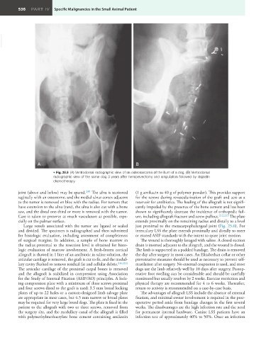
• Fig. 25.5 (A) Ventrodorsal radiographic view of an osteosarcoma of the ilium of a dog. (B) Ventrodorsal
radiographic view of the same dog 3 years after hemipelvectomy and amputation followed by cisplatin
chemotherapy.
joint (above and below) may be spared. 209 The ulna is sectioned (1 g amikacin to 40 g of polymer powder). This provides support
sagittally with an osteotome, and the medial ulnar cortex adjacent for the screws during revascularization of the graft and acts as a
to the tumor is removed en bloc with the radius. For tumors that reservoir for antibiotics. The healing of the allograft is not signifi-
have extension to the ulna (rare), the ulna is also cut with a bone cantly impeded by the presence of the bone cement and has been
saw, and the distal one-third or more is removed with the tumor. shown to significantly decrease the incidence of orthopedic fail-
Care is taken to preserve as much vasculature as possible, espe- ure, including allograft fracture and screw pullout. 212,213 The plate
cially on the palmar surface. extends proximally on the remaining radius and distally to a level
Large vessels associated with the tumor are ligated or sealed just proximal to the metacarpophalangeal joint (Fig. 25.6). For
and divided. The specimen is radiographed and then submitted intercalary LSS the plate extends proximally and distally to meet
for histologic evaluation, including assessment of completeness or exceed ASIF standards with the intent to spare joint motion.
of surgical margins. In addition, a sample of bone marrow in The wound is thoroughly lavaged with saline. A closed suction
the radius proximal to the resection level is obtained for histo- drain is inserted adjacent to the allograft, and the wound is closed.
logic evaluation of marrow involvement. A fresh-frozen cortical The limb is supported in a padded bandage. The drain is removed
allograft is thawed in 1 liter of an antibiotic in saline solution, the the day after surgery in most cases. An Elizabethan collar or other
articular cartilage is removed, the graft is cut to fit, and the medul- preventative measures should be used as necessary to prevent self-
lary cavity flushed to remove residual fat and cellular debris. 210,211 mutilation after surgery. No external coaptation is used, and most
The articular cartilage of the proximal carpal bones is removed dogs use the limb relatively well by 10 days after surgery. Postop-
and the allograft is stabilized in compression using Association erative foot swelling can be considerable and should be carefully
for the Study of Internal Fixation (ASIF/AO) principles. A lock- monitored but usually resolves by 2 weeks. Exercise restriction and
ing compression plate with a minimum of three screws proximal physical therapy are recommended for 4 to 6 weeks. Thereafter,
and four screws distal to the graft is used; 3.5 mm broad locking return to activity is recommended on a case-by-case basis.
plates of up to 22 holes or a custom-designed limb salvage plate The advantages of allograft LSS include the absence of external
are appropriate in most cases, but 4.5 mm narrow or broad plates fixation, and minimal owner involvement is required in the post-
may be required for very large breed dogs. The plate is fixed in the operative period aside from bandage changes in the first several
patient to the allograft with two or three screws, removed from weeks. The disadvantages are the high infection rate and the need
the surgery site, and the medullary canal of the allograft is filled for permanent internal hardware. Canine LSS patients have an
with polymethylmethacrylate bone cement containing amikacin infection rate of approximately 40% to 50%. Once an infection