
Page 560 - Withrow and MacEwen's Small Animal Clinical Oncology, 6th Edition
P. 560
538 PART IV Specific Malignancies in the Small Animal Patient
VetBooks.ir
b
a
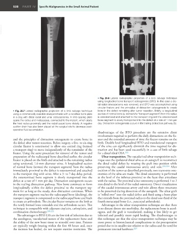
• Fig. 25.8 Lateral radiographic projection of a limb salvage technique
using longitudinal bone transport osteogenesis (BTO). In this case a dis-
tal radial osteosarcoma was removed, and BTO was accomplished using
circular fixators and the principles of distraction osteogenesis to create
• Fig. 25.7 Lateral radiographic projection of a limb salvage technique bone in the defect remaining after tumor resection. Briefly, a longitudinal
using a commercially available endoprosthesis with a modified bone plate section of normal bone (a, termed the “transport segment”) from the radius
in a dog with distal radial and ulnar osteosarcoma. A limb-sparing plate is osteotomized and attached to the transport ring and the osteotomized
spans the radius and metacarpus, connected to the implant, which abuts bone segment is slowly transported into the defect at a rate of 1 mm per
the host radius proximally and the radial carpal bone distally. A negative day. Distraction osteogenesis occurs in the trailing distraction pathway (b).
suction drain has also been placed at the surgical site to decrease post-
operative fluid accumulation.
disadvantages of the BTO procedure are the extensive client
involvement required to perform the daily distractions on the fix-
and the principles of distraction osteogenesis to create bone in ator and the extended amount of time the fixator remains on the
the defect after tumor resection. Before surgery, a five- to six-ring limb. Double level longitudinal BTO and translational transport
circular fixator is constructed to allow one central ring (termed of the ulna can significantly diminish the time required for dis-
a transport ring) to move independently of the remainder of the traction and has been used successfully in a case of limb salvage
fixator. Using the same procedure for removal of the tumor and for a distal tibial OSA. 222
preparation of the radiocarpal bone described earlier, the circular Ulnar transposition. The vascularized ulnar transposition tech-
fixator is placed on the limb and attached to the remaining radius nique uses the ipsilateral distal ulna as an autograft to reconstruct
using tensioned, 1.6-mm diameter wires. A longitudinal section the distal radial defect by rotating the graft into position while
of normal bone (termed the transport segment) from the radius preserving the caudal interosseous artery and vein. 223,224 After
immediately proximal to the defect is osteotomized and attached excision of the tumor as described previously, two transverse oste-
to the transport ring with wires. After a 3- to 7-day delay period, otomies of the ulna are made. The distal osteotomy is performed
the osteotomized bone segment is slowly transported into the at the level of the isthmus proximal to the facet that articulates
defect at a rate of 1 mm per day. Distraction osteogenesis occurs with the radius. The proximal ulnar osteotomy is performed 1 to 2
in the trailing distraction pathway. New bone continues to form mm distal to the level of the radial osteotomy. Direct visualization
longitudinally within the defect proximal to the transport seg- of the caudal interosseous artery and vein allows these structures
ment for as long as the steady, slow distraction continues. When to be preserved during dissection of the autograft. The ulnar graft
the transport segment reaches the radiocarpal bone (docking), the is “rolled over” into the radial defect and fixed using a bone plate
transport segment is compressed to the radiocarpal bone and heals that extends from the proximal radius to the distal one-third of the
to create an arthrodesis. The circular fixator remains on the limb as fourth metacarpal bone (i.e., pancarpal arthrodesis).
the newly formed bone remodels and the arthrodesis occurs. This Advantages to the ulnar transposition technique are that there
technique is compatible with adjuvant cisplatin, carboplatin, and is no distant donor site morbidity, the replacement bone is autol-
combination chemotherapy. 219,221 ogous, and the graft is vascularized, making it less likely to get
The advantages to BTO LSS are the low risk of infection due to infected and possibly more rapid healing. The disadvantages to
the autologous, vascularized nature of the replacement bone and this technique are that the ulnar transposition technique may be
the ability of the new bone tissue to remodel over time. Patients more prone to biomechanical complications in the postoperative
are typically weight-bearing within the first 48 hours and, once period due to its smaller size relative to the radius and the need for
the incision has healed, do not require exercise restriction. The permanent internal hardware. 223 