Page 620 - Withrow and MacEwen's Small Animal Clinical Oncology, 6th Edition
P. 620
598   PART IV    Specific Malignancies in the Small Animal Patient


         cases. 1,3,8,16   Although  rare,  thecomas  and  luteomas  are for  the
         most part considered benign. 8,16
            Although most sex cord stromal tumors are unilateral, bilat-
  VetBooks.ir  eral tumors are possible, especially among Sertoli–Leydig tumors.
                                                           8
         Concomitant cystic endometrial hyperplasia and cysts in the con-
         tralateral ovary appear common within this group. 8
            Solid, nest, cord, palisade, cystic, and spindle are the histo-
         logic patterns described for GTCT, and a mixture of these may
                                                          19
         exist within a single tumor. 8,19  GTCTs are generally vimentin,
         S-100,  and INH-α  positive, although one study has reported
               19
                          20
         INH-α–negative GTCT.  Variable expression of CK AE1/AE3,
                            19
                                                          20
                       22
         CK 7,  and ET-A  has been described. Moderate-to-strong intra-
              20
         cytoplasmic ET-1 immunoreactivity has been detected in 88% of
                                                          25
                     22
         canine GTCT,   whereas GTCT should be HBME-1 negative.
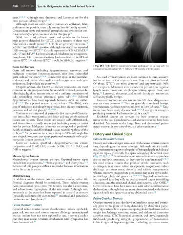
         Germ Cell Tumors                                      •  Fig. 27.1  Right lateral caudal abdominal radiograph of a dog with an
         Germ cell tumors, including dysgerminomas, teratomas, and   ovarian tumor. (Courtesy Dr. T. Schwarz, University of Edinburgh.)
         malignant teratomas (teratocarcinomas), arise from primordial
         germ cells of the ovary. 8,16,26  Concurrent cysts in the contralat-  Sex cord stromal tumors are most common in cats, account-
         eral ovary and uterine abnormalities such as pyometra and cystic   ing for at least half of reported cases. They are often unilateral.
         endometrial hyperplasia are common. 26                Of these, GTCTs are most common and approximately 50%
            Dysgerminomas, also known as ovarian seminomas, are most   are malignant. Metastatic sites include the peritoneum, regional
         common in this group and arise from undifferentiated germ cells.   lymph nodes, omentum, diaphragm, kidney, spleen, liver, and
         Histologically, these tumors consist of a uniform population of   lungs.  Luteomas, thecomas, and Sertoli–Leydig cell tumors are
                                                                    15
         cells  resembling ovarian primordial  germ cells. 3,8,16  Bilateral   rare and typically benign.
         dysgerminomas have been reported; however, most are unilat-  Germ cell tumors are also rare in cats. Of these, dysgermino-
         eral. 8,16,26  The reported metastatic rate is low (10%–30%), with   mas are most common.  They are generally considered benign,
                                                                                  15
         sites of metastasis including lymph nodes, liver, kidney, omentum,   yet metastasis has been reported in 20% to 33% of cases.  Tera-
                                                                                                            15
         pancreas, and adrenal glands. 3,8,9,26–28             tomas have been rarely documented. 15,30  A malignant estrogen-
            Teratomas are composed of germ cells that undergo differentia-  producing teratoma has been reported in a cat. 31
         tion into at least two germinal cell layers and any combination of   Epithelial tumors are perhaps the least common ovarian
         tissues can be seen. These tissues are usually well differentiated,   tumor in the cat. Cystadenomas and adenocarcinomas have been
         and tissues from virtually any organ (excluding ovary or testis)   described. Metastasis to the lungs, liver, and abdominal perito-
         may be present. Malignant teratomas are composed of predomi-  neum was seen in one case of ovarian adenocarcinoma.
                                                                                                          15
         nantly immature, undifferentiated tissues resembling those of the
                26
         embryo.  Metastasis has been noted in up to 50%. Although dis-  History and Clinical Signs
         tant visceral metastasis can occur, peritoneal metastasis with carci-
         nomatosis is most common. 8,9,26                      Canine Ovarian Tumors
            Germ cell tumors, specifically dysgerminomas, are vimen-  History and clinical signs associated with canine ovarian tumors
         tin positive and PLAP, CK7, desmin, S-100, CK AE1/AE3, and   vary, depending on the tissue of origin. Although initially insidi-
                      19
         INH-α negative.                                       ous, ovarian tumors grow to the point of being palpable and clinical
                                                               signs are typically referable to a space-occupying abdominal mass
         Mesenchymal Tumors                                    (Fig. 27.1). 9,32–34  Functional sex cord stromal tumors may produce
         Mesenchymal ovarian tumors are rare. Reported tumor types   one or multiple hormones, or they may be nonfunctional. 8,16,35
                                           16
         include hemangiosarcoma, 2,16  hemangioma,  and leiomyoma. 2,16    Sex cord stromal tumors that produce steroid hormones, such
         Behavior of this group is difficult to predict because information   as estrogen, may cause vulvar enlargement, sanguineous vulvar
         in the literature is sparse.                          discharge, persistent estrus, alopecia, and aplastic pancytopenia;
                                                               whereas excessive progesterone production may cause cystic endo-
         Miscellaneous                                         metrial hyperplasia and pyometra. 3,8,9,16,33  Hyperadrenocorticism
         In addition to the various primary ovarian tumors, other dif-  was reported in a dog with an ovarian-steroid tumor resembling
         ferential diagnoses should be considered. These include ovarian   a luteoma, and the associated clinical signs resolved after OHE.
                                                                                                                36
         cysts, paraovarian cysts, cystic rete tubules, vascular hamartomas,   Germ cell tumors have been associated with evidence of hormonal
         and adenomatous hyperplasia of the rete ovarii. Although rare,   dysfunction, although they are most often associated with clinical
         metastasis to the ovary has been reported in cases of mammary   signs referable to a space-occupying abdominal mass.
                                                                                                        26
         (especially inflammatory carcinoma),  intestinal and pancreatic
                                      29
                               16
         carcinoma, and lymphoma.                              Feline Ovarian Tumors
                                                               Ovarian tumors in cats also have an insidious onset and eventu-
         Feline Ovarian Tumors                                 ally grow to the point of being detectable by abdominal palpa-
         Reported feline ovarian tumor classifications include epithelial,   tion. Signs referable to a space-occupying abdominal mass such as
         germ cell, and sex cord stromal tumors. Although mesenchymal   weight loss, lethargy, vomiting, ascites, and abdominal distension
         ovarian tumors have not been reported in cats, it seems plausible   are often noted. GTCTs are most common, and they are generally
         that they may occur. Ovarian involvement with lymphoma has   functional, producing estrogen, progesterone, or testosterone.
         been documented. 15                                   Clinical  signs of hyperestrogenism, including persistent estrus,

