Page 621 - Withrow and MacEwen's Small Animal Clinical Oncology, 6th Edition
P. 621
CHAPTER 27  Tumors of the Female Reproductive System  599


                                                      0
  VetBooks.ir                                         1

                                                      2

                                                      3
                                                         x
                                                      4
                                                      5
                                                      6
            5.48cm
                                                      7                                                 50.0  m
                                 SAG   LT   OVARY
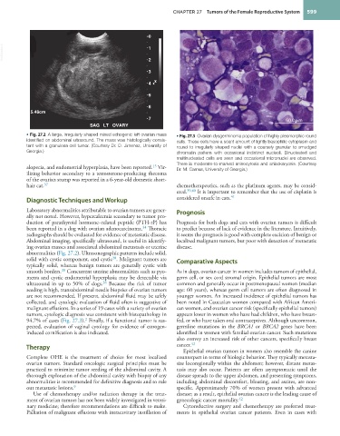
           • Fig. 27.2  A large, irregularly shaped mixed echogenic left ovarian mass   • Fig. 27.3  Ovarian dysgerminoma population of highly pleomorphic round
           identified on abdominal ultrasound. The mass was histologically consis-  cells. These cells have a scant amount of lightly basophilic cytoplasm and
           tent with a granulosa cell tumor. (Courtesy Dr. D. Jimenez, University of   round to irregularly shaped nuclei with a coarsely granular to smudged
           Georgia.)                                             chromatin pattern with occasional indistinct nucleoli. Binucleated and
                                                                 multinucleated cells are seen and occasional micronuclei are observed.
                                                                 There is moderate-to-marked anisocytosis and anisokaryosis. (Courtesy
                                                        15
           alopecia, and endometrial hyperplasia, have been reported.  Vir-  Dr. M. Camus, University of Georgia.)
           ilizing behavior secondary to a testosterone-producing thecoma
           of the ovarian stump was reported in a 6-year-old domestic short-
                 37
           hair cat.                                             chemotherapeutics, such as the platinum agents, may be consid-
                                                                 ered. 39,40  It is important to remember that the use of cisplatin is
                                                                                    41
           Diagnostic Techniques and Workup                      considered unsafe in cats.
           Laboratory abnormalities attributable to ovarian tumors are gener-  Prognosis
           ally not noted. However, hypercalcemia secondary to tumor pro-
           duction of parathyroid hormone–related peptide (PTH-rP) has   Prognosis for both dogs and cats with ovarian tumors is difficult
                                                    34
           been reported in a dog with ovarian adenocarcinoma.  Thoracic   to predict because of lack of evidence in the literature. Intuitively,
           radiographs should be evaluated for evidence of metastatic disease.   it seems the prognosis is good with complete excision of benign or
           Abdominal imaging, specifically ultrasound, is useful in identify-  localized malignant tumors, but poor with detection of metastatic
           ing ovarian masses and associated abdominal metastasis or uterine   disease.
           abnormalities (Fig. 27.2). Ultrasonographic patterns include solid,
           solid with cystic component, and cystic . Malignant tumors are   Comparative Aspects
                                         38
           typically solid, whereas benign tumors are generally cystic with
           smooth borders.  Concurrent uterine abnormalities such as pyo-  As in dogs, ovarian cancer in women includes tumors of epithelial,
                       38
           metra and cystic endometrial hyperplasia may be detectable via   germ cell, or sex cord stromal origin. Epithelial tumors are most
           ultrasound in up to 50% of dogs.  Because the risk of tumor   common and generally occur in postmenopausal women (median
                                      38
           seeding is high, transabdominal needle biopsies of ovarian tumors   age: 60 years), whereas germ cell tumors are often diagnosed in
           are not recommended. If present, abdominal fluid may be safely   younger women. An increased incidence of epithelial tumors has
           collected, and cytologic evaluation of fluid often is suggestive of   been noted in Caucasian women compared with African Ameri-
           malignant effusions. In a series of 19 cases with a variety of ovarian   can women, and ovarian cancer risk (specifically epithelial tumors)
           tumors, cytologic diagnosis was consistent with histopathology in   appears lower in women who have had children, who have breast-
           94.7% of cases (Fig. 27.3).  Finally, if a functional tumor is sus-  fed, or who have taken oral contraceptives. Although uncommon,
                                2
           pected, evaluation of vaginal cytology for evidence of estrogen-  germline mutations in the  BRCA1 or  BRCA2 genes have been
           induced cornification is also indicated.              identified in women with familial ovarian cancer. Such mutations
                                                                 also convey an increased risk of other cancers, specifically breast
           Therapy                                               cancer. 42
                                                                   Epithelial ovarian tumors in women also resemble the canine
           Complete OHE is the treatment of choice for most localized   counterpart in terms of biologic behavior. They typically metasta-
           ovarian tumors. Standard oncologic surgical principles must be   size locoregionally within the abdomen; however, distant metas-
           practiced to minimize tumor seeding of the abdominal cavity. A   tasis may also occur. Patients are often asymptomatic until the
           thorough exploration of the abdominal cavity with biopsy of any   disease spreads to the upper abdomen, and presenting symptoms,
           abnormalities is recommended for definitive diagnosis and to rule   including abdominal discomfort, bloating, and ascites, are non-
           out metastatic lesions. 9                             specific. Approximately 70% of women present with advanced
             Use of chemotherapy and/or radiation therapy in the treat-  disease; as a result, epithelial ovarian cancer is the leading cause of
           ment of ovarian tumors has not been widely investigated in veteri-  gynecologic cancer mortality. 42
           nary medicine; therefore recommendations are difficult to make.   Cytoreductive surgery and chemotherapy are preferred treat-
           Palliation of malignant effusions with intracavitary instillation of   ments in epithelial ovarian cancer patients. Even in cases with

