
Page 623 - Withrow and MacEwen's Small Animal Clinical Oncology, 6th Edition
P. 623
CHAPTER 27 Tumors of the Female Reproductive System 601
R
VetBooks.ir ∗
∗ ∗
∗
∗
UB
Sp
A C
∗
∗
∗ ∗ ∗
UB
UB
SP
B
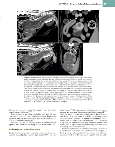
• Fig. 27.5 (A) Uterine body leiomyosarcoma. Computed tomographic sagittal reconstruction of the caudal
abdomen, postcontrast in delayed phase and displayed in soft tissue window. A uterine body mass (*)
extends 1 cm proximal to the external urethral orifice (white arrow). The mass is irregularly, mildly contrast
enhancing. (B) Uterine body leiomyosarcoma. Computed tomographic sagittal reconstruction (left) and
transverse plane (right) of the caudal abdomen, post-contrast in delayed phase and displayed in soft tis-
sue window. A uterine body mass (*) extends 1 cm proximal to the external urethral orifice (white arrow).
The mass is irregularly, mildly enhancing, a behavior consistent with the cystic spaces and dense cellular
arrangement observed on pathologic examination. The nodule on the splenic tail (calipers) was confirmed
to be lymphoid hyperplasia. SP: spleen, UB: urinary bladder. (C) Computed tomographic transverse recon-
struction of the caudal abdomen at the level of L4/5, postcontrast in delayed phase, soft tissue window. In
the region of the right ovary, a nodule (arrows) has a thin enhancing rim around a fluid dense center. These
findings correlate with histopathologic findings of a fluid to mucus-filled cyst partially lined by cells and
ovarian tissue, diagnosed as an interstitial cell tumor of an ovarian remnant. Cranial aspect of the uterine
body mass (*). (B Courtesy Dr. K. Gendron, University of Georgia.)
Lipomas often occur in younger dogs ranging in age from 1 to 8 vaginal tumors. 9,68 The most common malignant tumor is leiomyo-
years (mean age: 6.3 years). 46,68 sarcoma, and associated distant metastasis has been reported. 46,68
Malignant vaginal and vulvar tumors have been reported; how- Other less common tumor types include lipoma, fibrous histiocy-
ever, these appear to be more common in spayed female dogs. toma, benign melanoma, myxoma, myxofibroma, adenocarcinoma,
Primary clitoral carcinomas have been reported in a small number hemangiosarcoma, osteosarcoma, epidermoid carcinoma, and, in
of dogs, all of whom were spayed. 69 endemic areas, transmissible venereal tumor (TVT). 68,71 Carcinomas
Incidence rates are not available for the cat. Leiomyomas are arising from the bladder or urethra may manifest as vaginal masses
reported most commonly, occurring in older intact queens. 15,70 near the urethral papilla, and any skin tumor (e.g., mast cell tumors)
may develop on the labia of the vulva. 68
Pathology and Natural Behavior Macroscopically, tumors of the vestibule or vagina are described
9
as extraluminal or intraluminal. Extraluminal tumors appear as
Benign smooth muscle tumors, including leiomyoma, fibroleiomy- slowly growing, well-encapsulated, perineal masses. Intraluminal
oma, fibroma, and polyps, comprise approximately 83% of reported tumors are attached to the vestibular or vaginal wall by a pedicle,