
Page 177 - Anatomy and Physiology of Farm Animals, 8th Edition
P. 177
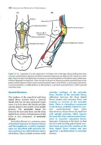
162 / Anatomy and Physiology of Farm Animals

                 (A)                                (B)
  VetBooks.ir          a       Palmar I.


                                          Suspensory I.

                                 Cruciate sesamoidean I.
                    e     e                              a                Palmar annular I.
                                                                e
                                     Extensor slip of
                                       suspensory I.
                                Oblique sesamoidean I.

                                                     b
                   b
                               Straight sesamoidean I.                 Digital flexor tendons


                       c

                       f                        c


                                                  f
                                          d
                       d


          Figure 8-11.  Ligaments (l.) in the equine foot. (A) Palmar view of the digit. Flexor tendons have been
          removed, and the palmar ligament and distal sesamoidean ligaments are depicted. (B) Lateral view of the
          digit. Deep and superficial digital flexor tendons are seen passing distad on the palmar aspect (distal sesa-
          moidean ligaments not depicted). These are held in the groove between proximal sesamoid bones by the
          palmar annular ligament which wraps around the palmar aspect of the fetlock. a, metacarpus/metatarsus;
          b, proximal phalanx; c, middle phalanx; d, distal phalanx; e, proximal sesamoid bones; f, distal sesamoid
          (navicular) bone.

          Synovial Structures                     articular cartilages of the navicular
                                                  bone, bursitis of the navicular bursa,
          The tendons of the superficial and deep   adhesions between the deep digital
          digital flexor muscles share a synovial   flexor tendon and navicular bone, and
          sheath that has its most proximal extent   erosions  or necrosis  of the navicular
          some 5 to 8 cm above the fetlock and that   bone. There is a hereditary component
          extends distad to the middle of the middle   to the disease, probably related to a cer-
          phalanx. The  navicular bursa lies      tain   conformational    type,   often
          between the navicular bone and the deep   described as a heavy horse on small feet
          digital flexor tendon. Inflammation of this   with upright pasterns, which exposes
          bursa is one component of  navicular    the navicular bone and associated struc-
          disease.                                tures to excessive concussive forces.
            Navicular disease is a common cause   Improper trimming of the hoof, so that
          of forelimb lameness in Quarter Horses   the toe is left too long and/or heels over-
          and Thoroughbreds. A variety of pathol-  shortened, increases the tension on the
          ogies are described with navicular dis-  deep digital flexor tendon and may
          ease, and not every affected horse shows   aggravate a predisposition to navicular
          all of them. These include erosion of the   disease.