
Page 181 - Anatomy and Physiology of Farm Animals, 8th Edition
P. 181
166 / Anatomy and Physiology of Farm Animals
(A) (B) (C) (D)
VetBooks.ir
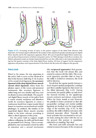
Figure 8-13. Increasing severity of injury to the passive support of the distal limb (thoracic limb
depicted). (A) Normal support afforded by the tendons of the superficial (purple) and deep (green) digital
flexor muscles and by the suspensory apparatus (blue). (B) Loss of superficial digital flexor tendon sup-
port produces hyperextension of fetlock and pastern joints. (C) With damage to both flexor tendons, the
fetlock and pastern joints are further hyperextended but now the coffin joint is also hyperextended, hav-
ing lost the passive restraint of the deep digital flexor tendon. (D) Loss of support via the suspensory
apparatus will produce such hyperextension of the fetlock that the joint will collapse to the ground.
Pelvic Limb (the reciprocal apparatus) that guaran-
tees that the hock will always flex and
Distal to the tarsus, the stay apparatus in extend in unison with the stifle. The recip-
the pelvic limb is more or less identical to rocal apparatus provides that as long as
that of the thoracic limb (Fig. 8‐14). In place the stifle is locked in extension, the hock
of the carpal check ligament, the accessory will be likewise.
ligament of the deep digital flexor muscle Recall that the equine patella features a
(tarsal check ligament) arises from the hook‐shaped medial parapatellar cartilage
plantar aspect of the tarsus and proximal and three patellar ligaments that insert on
metatarsus; this accessory ligament is the tibial tuberosity (Fig. 6‐12). During
rather long and very slender and may even movement, the patella glides up and down
be absent in some individuals. The superfi- the femoral trochlea as the quadriceps
cial digital flexor muscle of the pelvic limb muscle contracts and relaxes, respectively.
is nearly entirely tendinous and therefore When the horse stands at rest, however,
needs no accessory ligament to create a the patella is drawn proximad so that the
continuous band from origin (caudal distal parapatellar cartilage and medial patellar
femur) to insertion on proximal and middle ligament are hooked over the large medial
phalanges. In addition, the tendon of the ridge of the trochlea (Fig. 8‐15). In this
superficial digital flexor muscle attaches position, the stifle is held in extension with
firmly to the tuber calcanei, providing in minimal muscular effort. Typically, the
effect a check ligament at this point. horse activates this mechanism in one pel-
For the pelvic limb to support weight vic limb, resting the other with the toe on
without collapsing, the stifle and hock must the ground; this posture is sometimes
be prevented from flexing. This is accom- referred to as standing “hip‐shot.”
plished by one mechanism to lock the stifle The hock and stifle normally flex and
in extension and a second mechanism extend in unison because of the reciprocal