
Page 209 - Anatomy and Physiology of Farm Animals, 8th Edition
P. 209
194 / Anatomy and Physiology of Farm Animals
types of glia provide functions such as: (A) Schwann cells (B)
acting as immune cells; phagocytosing cel-
VetBooks.ir lular debris after trauma; regulating the
extracellular fluid environment; providing
myelination of axons; and many others
related to the normal function of nervous
tissue.
Nerve fibers may be myelinated or
unmyelinated. Myelinated fibers are
surrounded by a white sheath of fatty mate-
rial, or myelin. The myelin sheath actually
consists of many layers of cell membrane of
a specialized glial cell wrapped around
axons so that in cross‐section the myelin
sheath resembles a slice of jelly roll. In the
PNS, the myelinating cell is the Schwann
cell (neurolemmocyte), whereas in the
CNS, the oligodendrocyte fulfills this
function. Unmyelinated nerve fibers are
not exposed directly to the extracellular
fluid; rather, they are simply invaginated Myelin sheath
into the cell membrane of an adjacent glial
cell so that the cells surrounds the axon.
Axons covered in this way are not myeli-
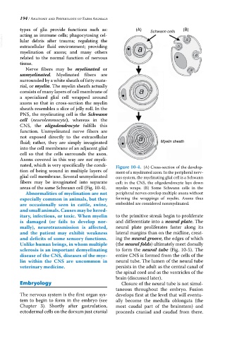
nated, which is very specifically the condi- Figure 10-4. (A) Cross‐section of the develop-
tion of being wound in multiple layers of ment of a myelinated axon. In the peripheral nerv-
glial cell membrane. Several unmyelinated ous system, the myelinating glial cell is a Schwann
fibers may be invaginated into separate cell; in the CNS, the oligodendrocyte lays down
areas of the same Schwann cell (Fig. 10‐4). myelin wraps. (B) Some Schwann cells in the
Abnormalities of myelination are not peripheral nerves envelop multiple axons without
especially common in animals, but they forming the wrappings of myelin. Axons thus
are occasionally seen in cattle, swine, embedded are considered nonmyelinated.
and small animals. Causes may be hered-
itary, infectious, or toxic. When myelin to the primitive streak begin to proliferate
is damaged (or fails to develop nor- and differentiate into a neural plate. The
mally), neurotransmission is affected, neural plate proliferates faster along its
and the patient may exhibit weakness lateral margins than on the midline, creat-
and deficits of some sensory functions. ing the neural groove, the edges of which
Unlike human beings, in whom multiple (the neural folds) ultimately meet dorsally
sclerosis is an important demyelinating to form the neural tube (Fig. 10‐5). The
disease of the CNS, diseases of the mye- entire CNS is formed from the cells of the
lin within the CNS are uncommon in neural tube. The lumen of the neural tube
veterinary medicine. persists in the adult as the central canal of
the spinal cord and as the ventricles of the
brain (discussed later).
Embryology Closure of the neural tube is not simul-
taneous throughout the embryo. Fusion
The nervous system is the first organ sys- develops first at the level that will eventu-
tem to begin to form in the embryo (see ally become the medulla oblongata (the
Chapter 3). Shortly after gastrulation, most caudal part of the brainstem) and
ectodermal cells on the dorsum just cranial proceeds craniad and caudad from there.