
Page 1211 - Veterinary Immunology, 10th Edition
P. 1211
VetBooks.ir
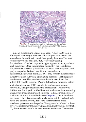
FIG. 37.1 A lymphocytic nodule in the thyroid of a dog suffering
from autoimmune thyroiditis. Original magnification ×100. (Courtesy
Dr. G. Stoica.)
In dogs, clinical signs appear after about 75% of the thyroid is
destroyed. These signs are those of hypothyroidism; that is, the
animals are fat and inactive and show patchy hair loss. The most
common problems are a dry, dull, coarse coat; scaling;
hypotrichosis; slow hair regrowth; hyperpigmentation; myxedema;
and pyoderma. Other signs include myopathy, hyperlipidemia,
hypothermia, anestrus, galactorrhea, diarrhea or constipation, and
polyneuropathy. Tests of thyroid function such as a
radioimmunoassay for plasma T or T only confirm the existence of
4
3
hypothyroidism. A thyroid-stimulating hormone (TSH) response
test is more useful because it can confirm the inability of the
affected thyroid to respond. (Plasma T levels are measured before
4
and after injection of TSH.) In order to confirm autoimmune
thyroiditis, a biopsy must show the characteristic lymphocytic
infiltration. Antithyroid antibodies must be detected in serum using
an enzyme-linked immunosorbent assay (ELISA), immunoblot, or
an indirect fluorescent antibody test (Chapter 42). As pointed out
previously, there is poor correlation between antithyroid antibody
titers and disease severity, reflecting the importance of cell-
mediated processes in this species. Management of affected animals
involves replacement therapy with sodium levothyroxine (synthetic
T ). Improvement should be seen within 4 to 6 weeks. There is no
4
1211