
Page 1228 - Veterinary Immunology, 10th Edition
P. 1228
VetBooks.ir
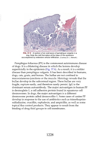
FIG. 37.5 A section of an oral lesion of pemphigus vulgaris in a
dog. Note the cleft formation at the base of the epidermis
accompanied by extensive cellular infiltration. (Courtesy Dr. J. Mansell.)
Pemphigus foliaceus (PF) is the commonest autoimmune disease
of dogs. It is a blistering disease in which the lesions develop
superficially in the epidermis (Fig. 37.6). As a result, it is a milder
disease than pemphigus vulgaris. It has been described in humans,
dogs, cats, goats, and horses. The bullae are not confined to
mucocutaneous junctions or the muzzle. Histology reveals that the
bullae develop in the subcorneal region. These bullae are very
fragile, rupture easily, and therefore rarely persist. IgG is the
dominant serum autoantibody. The major autoantigen in human PF
is desmoglein-1, a cell adhesion protein found in squamous cell
desmosomes. In dogs, the major autoantigen is a different
desmosome protein called desmocollin-1. Some cases of canine PF
develop in response to the use of antibiotics such as trimethoprim-
sulfadiazine, oxacillin, cephalexin, and ampicillin, as well as some
topical flea control products. They appear to result from the
binding of drug thiol groups to cell membranes.
1228