
Page 112 - Veterinary Laser Therapy in Small Animal Practice
P. 112
98 Veterinary Laser Therapy in Small Animal Practice
Case no. 12
M., canine, 6 years old, Labrador, FS, 27 kg
• Complaint: fistula.
• History:
• M. was referred for surgery for a chronic fistula in her caudal right flank, which had appeared for
the first time 2.5 years ago. It had undergone three previous surgeries in other practices. The fistula
would improve with steroid treatment, but recur after its withdrawal. Antibiotics did not seem to help.
Reopenings of the fistula were sometimes preceded by a clear vaginal discharge.
• A month ago, she had been treated for proximal ureteral stenosis and mild hydronephrosis, using
prazosin and tramadol, with a good response. The initial abdominal ultrasound had evidenced a
hyperechoic structure in the proximal ureter, but later on it could not be located and calculi/foreign
material had been ruled out.
• Urinary culture was negative. Blood culture showed leukocytosis with neutrophilia.
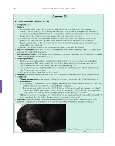
• Physical examination: open fistula in the caudal right flank, with purulent discharge (Fig. C12.1). Scars from
previous draining tracts and surgeries.
• Complementary tests: an MRI was performed before surgery, and a connection from the fistula to the caudal
area of the right kidney was evident (Fig. C12.2).
• Surgical procedure:
• First, an exploratory laparotomy was performed. Both ovarian and uterine pedicles had braided non-
absorbable suture. The fibrosis around the right pedicle had entrapped the proximal ureter. All non-
absorbable sutures were removed and the ureter was released (Fig. C12.3).
• The fistula was opened along the right flank and partially debrided along its main tract. Before closure,
two Penrose drains were placed.
• Culture of the fistula showed no bacterial growth.
• Diagnosis: chronic inflammatory fistulous tract due to inadequate use of non-absorbable suture material.
• Treatment:
• Wound management: regular cleaning. One of the drains was removed on day 3, the other on day 5.
• Laser therapy:
• In this case, LT was started 24 h after surgery. Treatment area on the flank was around 300–350 cm ,
2
and the initial was dose 3 J/cm , so a total of 1000 J was used.
2
• Treatments were performed on days 1, 3, 5, 7, 10, and 14 (suture removal) after surgery. From day 5
onward, 5 J/cm was used, with a total of 1500 J per treatment. On day 14, contact mode was used,
2
2
using a 5 cm spot size and therefore a slightly higher power density (0.6 W/cm ).
2
• Others: marbofloxacin 2 mg/kg q24h × 3 days (until culture results available), tramadol 3 mg/kg q8h × 3
days, prednisone 1 mg/kg q24h × 5 days, and then progressively weaning off.
• Outcome: uneventful healing. Surgery resolved the origin of the problem and LT prevented many of the
potential complications of this case, such as seroma, recurrence of the fistula, delayed healing, etc.
Figure C12.1 Opening of the fistulous tract in the right
flank.
REDONDO PRINT (4-COL BLEED).indd 98 08/08/2019 09:48