
Page 107 - Veterinary Laser Therapy in Small Animal Practice
P. 107
Pointing light at soft tissue: clinical applications 93
Case no. 9
B., canine, 7 years old, Golden Retriever, MC, 32 kg
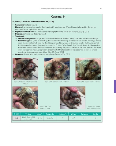
• Complaint: foot pad wound.
• History: B. underwent surgery for fistulous tract 3 months prior. Wound has not changed for 2 months
despite different topical treatments.
• Physical examination: 9 × 4 mm wound in the right hindlimb pad of the fourth digit (Fig. C9.1).
• Diagnosis: chronic non-healing wound.
• Treatment:
• Wound management: lavage with 0.05% chlorhexidine. Manuka honey ointment. Protective bandage.
• Laser therapy: 10 J/cm as a starting dose due to the chronicity and depth of the wound. Although it may
2
seem like a small defect, note the dead tissue around the wound, with excess keratin that is unattached
to the underlying tissue. Dose was increased to 15 J/cm after 1 week (6 × 5 mm). Again, in this case the
2
treatment area included the blood vessels running along the plantar surface of the paw. Before a decrease
in wound size was noted, progressive attachment of the keratin layer was observed as well as a more
reactive and vascularized wound bed (Figs C9.2 and C9.3).
• Outcome: closure after six treatments spread over 1 month (Fig. C9.5).
Figure C9.1 Before initial treatment. 9 Figure C9.2 After one week. 6 × 5 mm. Figure C9.3 Second week. 6 × 5 mm.
× 4 mm.
Figure C9.4 Third Figure C9.5 Fourth
week. 3 × 1 mm. week. Wound closure.
P (W) Tx time J/cm 2 Total J/Tx Spot (cm ) W/cm 2 Tx/week No. Tx
2
a
85 s (1.4 min)–128
3.5 10–15 300–450 5 0.7 2-1 6
s (2.1 min)
REDONDO PRINT (4-COL BLEED).indd 93 08/08/2019 09:47