
Page 106 - Veterinary Laser Therapy in Small Animal Practice
P. 106
92 Veterinary Laser Therapy in Small Animal Practice
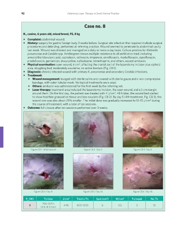
Case no. 8
K., canine, 6 years old, mixed breed, FS, 8 kg
• Complaint: abdominal wound.
• History: surgery for gastric foreign body 3 weeks before. Surgical site infection that required multiple surgical
procedures and debriding, performed at referring practice. Wound seemed to penetrate to abdominal cavity
last week. Wound was dressed and managed on a daily or twice a day basis. Culture positive for Klebsiella
pneumoniae and Candida spp. Antibiogram shows multiple resistance to all antibiotics tried, including
amoxicillin/clavulanic acid, cephalexin, cefovecin, imipenem, enrofloxacin, marbofloxacin, ciprofloxacin,
pradofloxacin, gentamicin, doxycycline, sulfadiazine, trimethoprim, and others, except amikacin.
2
• Physical examination: open wound, 6 cm , affecting the cranial part of the laparotomy incision plus xiphoid
area, sloughing bed, moderately exudative, no active borders (Fig. C8.1).
• Diagnosis: chronic infected wound with primary K. pneumoniae and secondary Candida infections.
• Treatment:
• Wound management: lavaged with sterile saline and covered with sterile gauze and a non-compressive
bandage, with outer tubular mesh. No topical treatments were used.
• Others: amikacin was administered for the first week by the referring vet.
• Laser therapy: treatment area included the laparotomy incision, the open wound, and a 5 cm margin
around them. On the first day, the patient was treated with 4 J/cm ; 48 h later, the wound bed started
2
to show healthier granulation tissue and less exudate (Fig. C8.2). By day 5 (4th treatment, Fig. C8.3), the
wound size was also about 25% smaller. The initial dose was gradually increased to 12–15 J/cm during
2
the course of treatment, with a total of ten sessions.
• Outcome: full closure after ten sessions performed over 3 weeks.
Figure C8.1 Initial wound. Figure C8.2 Day 2. Figure C8.3 Day 5.
Figure C8.4 Day 9. Figure C8.5 Day 14. Figure C8.6 Day 18.
2
P (W) Tx time J/cm 2 Total J/Tx Spot (cm ) W/cm 2 Tx/week No. Tx
a
266–500 s
3 4–15 800–1200 6 0.5 3 10
(4.4–8.3 min)
REDONDO PRINT (4-COL BLEED).indd 92 08/08/2019 09:47