
Page 103 - Veterinary Laser Therapy in Small Animal Practice
P. 103
Pointing light at soft tissue: clinical applications 89
Case no. 5
O., canine, 9 years old, mixed breed, MC, 20 kg
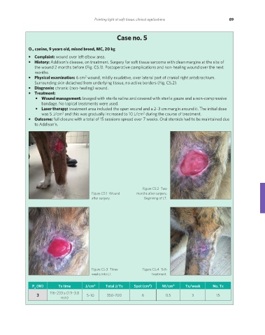
• Complaint: wound over left elbow area.
• History: Addison’s disease, on treatment. Surgery for soft tissue sarcoma with clean margins at the site of
the wound 2 months before (Fig. C5.1). Postoperative complications and non-healing wound over the next
months.
2
• Physical examination: 6 cm wound, mildly exudative, over lateral part of cranial right antebrachium.
Surrounding skin detached from underlying tissue, no active borders (Fig. C5.2).
• Diagnosis: chronic (non-healing) wound.
• Treatment:
• Wound management: lavaged with sterile saline and covered with sterile gauze and a non-compressive
bandage. No topical treatments were used.
• Laser therapy: treatment area included the open wound and a 2–3 cm margin around it. The initial dose
was 5 J/cm and this was gradually increased to 10 J/cm during the course of treatment.
2
2
• Outcome: full closure with a total of 15 sessions spread over 7 weeks. Oral steroids had to be maintained due
to Addison’s.
Figure C5.2 Two
Figure C5.1 Wound months after surgery.
after surgery. Beginning of LT.
Figure C5.3 Three Figure C5.4 15th
weeks into LT. treatment.
2
P (W) Tx time J/cm 2 Total J/Tx Spot (cm ) W/cm 2 Tx/week No. Tx
a
3 116–233 s (1.9–3.8 5–10 350–700 6 0.5 3 15
min)
REDONDO PRINT (4-COL BLEED).indd 89 08/08/2019 09:47