
Page 99 - Veterinary Laser Therapy in Small Animal Practice
P. 99
Pointing light at soft tissue: clinical applications 85
Case no. 2
J., canine, 15 years old, Belgian Shepherd, FS, 25 kg
• Complaint: wound on left ear.
• History: chronic wound had appeared for the first time 2 years previously. No conclusive diagnosis could
be reached, although vasculitis was suspected. Systemic infections such as leishmaniasis had been ruled
out. Several long courses of antibiotics and steroids had been used, with an initial good response, but the
wound would reopen as soon as medications were stopped. No response to local treatments. Last dose of
triamcinolone had been a week before the case was referred for LT.
• Physical examination: 2–3 cm in size (Fig. C2.1), almost full thickness wound on the distal third of left ear
2
pinna. No granulation present. No exudates.
• Diagnosis: chronic non-healing wound due to suspected vasculitis.
• Treatment:
• Wound management: lavage with saline if any debris present.
• Laser therapy:
• The initial treatment used 4–5 J/cm , but after three treatments there was no improvement (Fig. C2.2
2
2
and C2.3), so dose was increased to 8 J/cm .
• After day 10 (6th treatment), the change was very subtle so dose was again increased to 12 J/cm ,
2
which seemed to be of significant help; by session 12 the wound was no smaller but the bed was filled
2
with healthy granulation tissue and dose was increased to 16 J/cm .
• One month after beginning LT, active epithelialization was evident at the edges (Fig. C2.5). Dose was
increased to 20 J/cm .
2
• On day 40, dose was increased to 25 J/cm . By day 52, only an 8 × 1 mm defect remained.
2
2
2
• In this case, 2 min of time off were taken between each 4–5 J/cm . So eventually, when 25 J/cm was
being delivered, the session length was around 15 minutes long: around 1 min on, 2 min off.
• Others: no other local nor systemic medications were used during the course of laser treatments.
• Outcome: full closure in 2 months (Fig. C2.8) with 22 sessions. This patient would be considered a weak
reactor, since it took 2 months to resolve (although the wound was present for 2 years) and needed very high
doses of LT. Three more weekly treatments were performed to prevent reopening. The steroid treatment
could have contributed to the slow initial response.
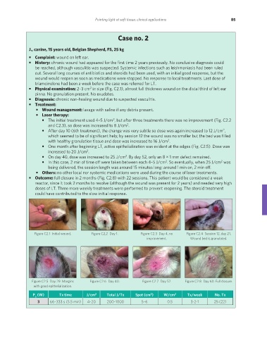
Figure C2.1 Initial wound. Figure C2.2 Day 1. Figure C2.3 Day 4, no Figure C2.4 Session 12, day 21.
improvement. Wound bed is granulated.
Figure C2.5 Day 29. Margins Figure C2.6 Day 40. Figure C2.7 Day 52. Figure C2.8 Day 60. Full closure.
with good epithelialization.
2
P (W) Tx time J/cm 2 Total J/Tx Spot (cm ) W/cm 2 Tx/week No. Tx
a
3 66–333 s (5.5 min) 4–20 200–1000 5–6 0.5 3-2-1 25 (22)
REDONDO PRINT (4-COL BLEED).indd 85 08/08/2019 09:47