
Page 162 - Veterinary Laser Therapy in Small Animal Practice
P. 162
148 Veterinary Laser Therapy in Small Animal Practice
Case no. 22
T., canine, 6 years old, Boxer, MC, 37 kg
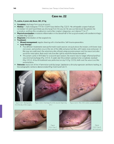
• Complaint: discharge from surgical wound.
• History: T. had undergone TTA for CCLR 11 days before (Fig. C22.1). His orthopedic surgeon had just
rechecked him and found there was discharge from the wound. He was worried about the patient, the
procedure, and how this complication could affect implant integration, and referred T. for LT.
• Physical examination: moderate inflammation in the dorsal half of the surgical wound, with serohemorrhagic
exudate (Fig. C22.2).
• Diagnosis: inflammation of the surgical site.
• Treatment:
• Wound management: regular cleaning with chlorhexidine. Self-trauma prevention.
• Laser therapy:
• Two different treatments were performed in each session: one just above the incision, with lower dose
and power, and another around the rest of the stifle and proximal tibia, with higher dose and power. In
this way we could reach the osteotomy site without using excessive power over the wound and also
avoid the metal plate, that would not allow the light to reach the tissues beneath it.
• The patient came in 48 h later for a second laser session; the wound was no longer inflamed and had
very minimal discharge (Fig. C22.3). A week later the problem seemed to be completely resolved
(Fig. C22.4). A fourth treatment was performed on day 9 (Fig. C22.5), both over the wound and the
proximal tibia.
• Outcome: resolution of the inflammation and discharge. Satisfactory clinical progression and bone healing, as
the radiographic rechecks demonstrated (Figs C22.6 and C22.7).
Figure C22.1 Radiography Figure C22.2 Discharge from the wound. Beginning Figure C22.3 After 48 h, before second treatment.
from the day of the surgery. of LT.
Figure Figure
C22.4 Day 7, C22.5 Day 9,
third treatment. last treatment.
REDONDO PRINT (4-COL BLEED).indd 148 08/08/2019 09:48