
Page 107 - Veterinary Histology of Domestic Mammals and Birds, 5th Edition
P. 107
Connective and supportive tissues (textus connectivus) 89
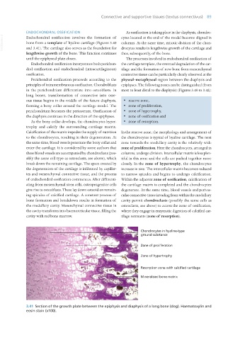
ENDOCHONDRAL OSSIFICATION As ossification is taking place in the diaphysis, chondro-
VetBooks.ir Endochondral ossification involves the formation of cytes located at the end of the model become aligned in
bone from a template of hyaline cartilage (Figures 3.40 columns. At the same time, mitotic division of the chon-
and 3.41). The cartilage also serves as the foundation for drocytes results in lengthwise growth of the cartilage and
lengthwise growth of the bone. This function continues thus, subsequently, of the bone.
until the epiphyseal plate closes. The processes involved in endochondral ossification of
Endochondral ossification incorporates both perichon- the cartilage template, the eventual degradation of the car-
dral ossification and endochondral (intracartilaginous) tilage and the formation of new bone from mesenchymal
ossification. connective tissue can be particularly clearly observed at the
Perichondral ossification proceeds according to the physeal–metaphyseal region between the diaphysis and
principles of intramembranous ossification. Chondroblasts epiphyses. The following zones can be distinguished (from
in the perichondrium differentiate into osteoblasts. In most to least distal to the diaphysis) (Figures 3.40 to 3.42):
long bones, transformation of connective into osse-
ous tissue begins in the middle of the future diaphysis, · reserve zone,
forming a bony collar around the cartilage model. The · zone of proliferation,
perichondrium becomes the periosteum. Ossification of · zone of hypertrophy,
the diaphysis continues in the direction of the epiphyses. · zone of ossification and
As the bony collar develops, the chondrocytes hyper- · zone of resorption.
trophy and calcify the surrounding cartilage matrix.
Calcification of the matrix impedes the supply of nutrition In the reserve zone, the morphology and arrangement of
to the chondrocytes, resulting in their degeneration. At the chondrocytes is typical of hyaline cartilage. The next
the same time, blood vessels penetrate the bony collar and zone towards the medullary cavity is the relatively wide
enter the cartilage. It is considered by some authors that zone of proliferation. Here the chondrocytes, arranged in
these blood vessels are accompanied by chondroclasts (pos- columns, undergo division. Intercellular matrix is less plen-
sibly the same cell type as osteoclasts, see above), which tiful in this zone and the cells are packed together more
break down the remaining cartilage. The space created by closely. In the zone of hypertrophy, the chondrocytes
the degeneration of the cartilage is infiltrated by capillar- increase in size. The intercellular matrix becomes reduced
ies and mesenchymal connective tissue, and the process to narrow spicules and begins to undergo calcification.
of endochondral ossification commences. After differenti- Within the adjacent zone of ossification, calcification of
ating from mesenchymal stem cells, osteoprogenitor cells the cartilage matrix is completed and the chondrocytes
give rise to osteoblasts. These lay down osteoid on remain- degenerate. At the same time, blood vessels and perivas-
ing spicules of calcified cartilage. A constant process of cular connective tissue invading from within the medullary
bone formation and breakdown results in formation of cavity permit chondroclasts (possibly the same cells as
the medullary cavity. Mesenchymal connective tissue in osteoclasts, see above) to access the zone of ossification,
the cavity transforms into haemoreticular tissue, filling the where they engage in enzymatic digestion of calcified car-
cavity with red bone marrow. tilage remnants (zone of resorption).
Chondrocytes in hyaline-type
ground substance
Zone of proliferation
Zone of hypertrophy
Resorption zone with calcified cartilage
Mineralised bone matrix
3.41 Section of the growth plate between the epiphysis and diaphysis of a long bone (dog). Haematoxylin and
eosin stain (x100).
Vet Histology.indb 89 16/07/2019 14:56