
Page 114 - Veterinary Histology of Domestic Mammals and Birds, 5th Edition
P. 114
96 Veterinary Histology of Domestic Mammals and Birds
cellular compartment. The tubules of the T system form characteristic banding pattern (light and dark) arising
VetBooks.ir ring-like anastomoses yet are separated from the terminal from the ordered, parallel arrangement of their compo-
cisternae of the sarcoplasmic reticulum by a space. The nent myofilaments (Figures 4.7 to 4.14). The bands can be
region in which two terminal cisternae of the sarcoplas-
distinguished on the basis of their refractive properties:
mic reticulum lie adjacent to a tubule of the T system is
referred to as a triad (Figure 4.7). · isotropic, monorefringent (= light) I bands and
The T system enables rapid transmission of the nerve · anisotropic, birefringent (= dark) A bands.
impulse (depolarisation with influx of sodium) from the cell
membrane to the interior of the cell and facilitates coordi- These bands are bisected by transverse bands of different
nated contraction of the whole muscle fibre. At the triads, the density:
impulse is transmitted to the sarcoplasmic reticu-
2+
2+
lum, from which Ca is released. Ca thus acts as · The I band is bisected by a dense intermediate disc
a mediator between electrical stimulation at the cell (‘Zwischenscheibe’) termed the Z line.
membrane and contraction of the myofibrils within the · The A band is bisected by a less dense region (H band)
cell. which in turn is bisected by the narrow, dense M line.
A single muscle fibre is estimated to contain around
1000 myofibrils. In longitudinal section, these exhibit a
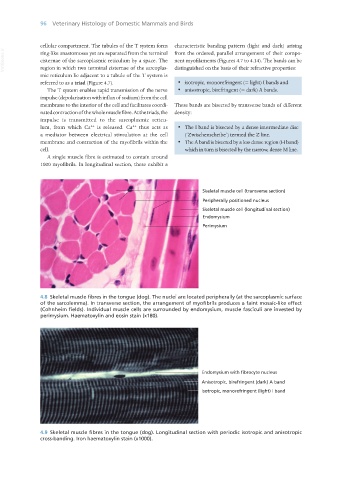
4.8 Skeletal muscle fibres in the tongue (dog). The nuclei are located peripherally (at the sarcoplasmic surface
of the sarcolemma). In transverse section, the arrangement of myofibrils produces a faint mosaic-like effect
(Cohnheim fields). Individual muscle cells are surrounded by endomysium, muscle fasciculi are invested by
perimysium. Haematoxylin and eosin stain (x180).
Endomysium with fibrocyte nucleus
Anisotropic, birefringent (dark) A band
Isotropic, monorefringent (light) I band
4.9 Skeletal muscle fibres in the tongue (dog). Longitudinal section with periodic isotropic and anisotropic
cross-banding. Iron haematoxylin stain (x1000).
Vet Histology.indb 96 16/07/2019 14:56