
Page 133 - Veterinary Histology of Domestic Mammals and Birds, 5th Edition
P. 133
Nervous tissue (textus nervosus) 115
nutrition and metabolic support to neurons and facilita- · neuronal regeneration, in which they act as a physi-
VetBooks.ir tion of the conduction of nerve impulses by the formation · contribution to the blood–brain barrier and other
cal guide,
of neural sheaths. In some cases, glial cells contribute to
innate immunity as specialised cells of the mononuclear
phagocyte system. interfaces in the central nervous system and
Most nerve cells are closely related, both structurally · capacity for phagocytosis (certain cells).
and functionally, to glial cells (neuroglia), with which they
share a common embryonic origin. Specific functions Based on structural and functional criteria (Table 5.1), glial
performed by glial cells include: cells are divided into:
· physical support by occupying the spaces between · glial cells of the central nervous system:
perikarya, dendrites and axons, thus contributing to − ependymal cells,
the organisation and spatial separation of neurons, − astrocytes,
· metabolic support and exchange of substances − oligodendrocytes and
between the nerve cells and capillaries, − microglia (Hortega cells) (Figures 5.21 to 5.25);
· formation of the sheath of myelinated and unmy- · glial cells of the peripheral nervous system:
elinated nerve fibres, thus influencing the speed of − Schwann cells (neurolemmocytes) and
nerve impulse conduction, − satellite cells (amphicytes).
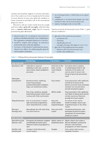
Table 5.1 Distinguishing microscopic features of neuroglia.
Morphology Occurrence Function
Cells of the CNS
Ependymal cells Cuboidal to columnar Ventricles of the Formation and circulation
epithelium with cilia, round to brain, central of cerebrospinal fluid in the
oval nucleus, basal cytoplasmic canal of spinal ventricles of the brain and
processes cord central canal of spinal cord,
lining of ventricles and central
canal
Astrocytes:
protoplasmic Numerous short, radiating, Grey matter Close association with capillaries,
branching processes, contribute to blood–brain
prominent/abundant cytoplasm, barrier, surround nerve processes,
large, oval euchromatic nucleus maintain ion concentration,
provide structural support
fibrous Few, long, radiating, sparsely White matter Close association with capillaries,
branched processes, small round contribute to blood–brain barrier,
nucleus with little euchromatin contain numerous filaments,
provide structural support
Oligodendrocytes Few or no processes in routine Grey and Formation of the myelin
light microscopic preparations, white matter sheath (multiple neurons per
small, heterochromatic nucleus (oligodendroglia) oligodendrocyte) in the white
matter
Microglia Macrophage-like cells originating Grey and white Amoeboid phagocytes, well-
(Hortega cells) from bone marrow matter developed lysosomal system,
component of mononuclear
phagocytic system
Cells of the PNS
Neurolemmocytes Elongated cell extension wrapped Myelin sheath of Myelin sheath and nodes of
(Schwann cells) concentrically around an axon, peripheral axons Ranvier between consecutive
flattened nucleus lying adjacent to Schwann cells facilitate
axon propagation of nerve impulses
Satellite cells Compact, heterochromatic, Ganglia Close contact with neurons,
(amphicytes) round nucleus metabolic functions
Vet Histology.indb 115 16/07/2019 14:57