
Page 131 - Veterinary Histology of Domestic Mammals and Birds, 5th Edition
P. 131
Nervous tissue (textus nervosus) 113
as Schmidt–Lanterman clefts, can be clearly seen in lon- the surface of the axolemma. The spread of the impulse is
VetBooks.ir gitudinal sections of nerve fibres. It is thought they may slower due to the absence of nodes of Ranvier (and thus of
saltatory conduction) and inferior conductivity of unmy-
facilitate deformation of the nerve within the tissue.
Unmyelinated nerve fibres elinated fibres.
In the Erlanger–Gasser system, nerve fibres are classi-
In unmyelinated nerve fibres, the Schwann cell surrounds fied as A, B or C on the basis of diameter and conduction
the axon without subsequent wrapping by the mesaxon. speed. A fibres are myelinated, rapidly conducting fibres
In contrast to myelinated peripheral fibres, in which a that are predominantly associated with muscle fibres,
Schwann cell is associated with only one axon, several muscle spindles or skin. They may be efferent or afferent.
axons are usually surrounded by the same Schwann cell in A fibres may be further subdivided into subgroups by their
unmyelinated fibres (Figure 5.15). Neighbouring Schwann diameter and conduction speed.
cells are closely apposed, such that nodes of Ranvier are Aα fibres (efferent and afferent innervation of muscle)
not observed. In the central nervous system, unmyelinated are the thickest and fastest-conducting fibres (10–20 μm
axons often lie immediately adjacent to other neurons or and 60–120 m/s). Below these in speed and diameter are
glial cell processes. the Aβ fibres (afferent fibres, touch and pressure) which
measure 7–15 μm in diameter and conduct at 40–90 m/s.
Generation and conduction of nerve stimuli With a diameter of 4–8 μm, Aγ fibres (efferent to muscle
The initiation and transmission of an electrical impulse spindles) are also relatively slow (30–45 m/s). Aδ fibres (3–5
occurs at the plasmalemma (axolemma) of the neuron. μm) are slow-conducting fibres (5–25 m/s) involved in the
Central to this process is a change in the voltage across monitoring of pain and temperature.
the membrane, resulting from the movement of particu- B fibres (1–3 μm) are myelinated fibres that conduct
+
+
–
lar inorganic ions (Na , K , Cl and Ca ) through specific the action potentials of preganglionic autonomic fibres at
2+
channels within membrane proteins (ion channels). a moderate speed of 3–15 m/s.
Opening or closing of the ion channels alters the dis- C fibres are thin, unmyelinated nerve fibres (0.3–1 μm)
tribution of charge across the membrane and thus the that conduct very slowly (0.5–2 m/s). They serve to convey
membrane potential (voltage). An action potential is impulses in postganglionic autonomic nerve fibres.
generated when Na channels open in response to a depo-
+
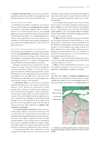
larising stimulus, with a subsequent increase in membrane Nerves
+
+
permeability to Na and influx of Na ions into the cell. The term ‘nerve’ applies to a bundle of peripheral nerve
+
This influx exceeds the efflux of K ions, altering the rest- fibres that are combined by connective tissue into single,
ing potential to create an action potential. variably sized entities. Nerves containing fibres with vary-
In myelinated fibres, the alteration of membrane ing characteristics are referred to as mixed nerves. In
potential can only occur at the nodes of Ranvier. The
+
majority of Na channels are concentrated in these
regions, with a density of several thousand channels per Epineurium
2
μm . The internode, the segment of the nerve fibre sur-
Adipose tissue
rounded by the myelin sheath, is virtually devoid of Na
+
channels. The myelin sheath contributes significantly to Perineurium
nerve conduction by insulating the cell and preventing
leakage of current. Consequently, the nerve impulses that Endoneurium
give rise to the action potential ‘jump’ from one node of
Ranvier to the next. This is referred to as saltatory (dis-
continuous) conduction.
The speed of impulse conduction within a neuron is
constant and is related to the structure of the axon. This
is accelerated by the myelin sheath, which enables the
rapid spread of action potentials from one node of Ranvier
to the next. Thus, saltatory impulse transmission through
myelinated fibres is faster than the type of transmission
that occurs in unmyelinated fibres. In addition, myeli-
nated fibres require less energy as the processes involved
in impulse conduction are limited to the nodes of Ranvier. 5.17 Mixed nerve fibre bundle with connective tissue
In unmyelinated fibres, the conduction of nerve investments (dog). Goldner’s Masson trichrome stain
impulses to the synapse occurs in a continuous wave over (x120).
Vet Histology.indb 113 16/07/2019 14:57