
Page 147 - Veterinary Histology of Domestic Mammals and Birds, 5th Edition
P. 147
Circulatory system (systema cardiovasculare et Iymphovasculare) 129
Arteriovenous specialisations
VetBooks.ir Structures involved in regulating blood flow are present in
most organs of the body. These are associated with arteri-
oles and venules, particularly at sites of vessel branching,
and in pre- and postcapillary segments. They include:
· barrier arteries ('polster arteries'),
· throttle veins ('sphincter veins'),
· arteriovenous anastomoses
− bridge form
− coiled (glomus) form.
In barrier arteries, longitudinally oriented smooth
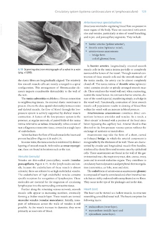
6.19 Scanning electron micrograph of a valve in a vein muscle cells in the tunica intima partially or completely
(pig; x1200). surround the lumen of the vessel. Through maximal con-
traction of these muscle cells and the smooth muscle of
the elastic fibres are longitudinally aligned. The relatively the tunica media, the artery can be almost completely
few smooth muscle cells are mostly arranged in a spiral closed off. The tunica intima of throttle veins (sphincter
configuration. This arrangement of fibromuscular ele- veins) contains circular or spirally arranged smooth mus-
ments imparts considerable distensibility to the wall of cle. These reinforce the vessel wall and, when contracting,
the vein. restrict the vessel lumen. In contrast to barrier arteries, clo-
The tunica adventitia establishes a fibrous connection sure of the vessel is partial, manifesting simply as a bulge in
to neighbouring tissues. An external elastic membrane is the vessel wall. Functionally, contraction of these smooth
present. Due to the close spatial relationship between veins muscle cell populations results in slowing of blood flow
and skeletal muscle, the flow of blood through the low- within the vessel with an increase in blood pressure.
pressure system is actively supported by skeletal muscle Arteriovenous anastomoses constitute direct con-
contraction. A feature of the low-pressure system is the nections between arterioles and venules. As a result, a
presence, at regular intervals, of paired folds of the tunica ‘short-circuit’ is formed with a portion of the local circu-
intima referred to as valves. Structurally, valves consist of lation bypassing the capillary bed. Arterial blood is thus
taut collagenous connective tissue, covered in a single layer diverted to the low-pressure venous system without the
of endothelium. exchange of nutrients or metabolites.
Valves facilitate the flow of blood towards the heart and Anastomoses may take the form of a short, curved
prevent backflow (Figures 6.18 and 6.19). or S-shaped bridge, in which the arterial component is
In some veins, the tunica media is reinforced by distinct recognisable by the thickness of its wall. These are charac-
layering of smooth muscle. Referred to as venae myotypi- terised by circular and longitudinal muscle fibre bundles,
cae, these are found in locations such as the teat. reinforced by elastic fibres and in some cases by epithelioid
cells. These anastomoses are found in the wall of the gas-
Venules (venula) trointestinal tract, the respiratory tract, skin, uterus, ovary,
Venules are thin-walled postcapillary vessels (venulae penis and in several endocrine organs. They contribute to
postcapillares; Figure 6.15). In the lymph nodes and ton- circulatory haemodynamics and peripheral thermoregula-
sils, because the endothelium of these vessels is almost tion (extremities, ear lobe, nose and skin).
cuboidal, these are referred to as high endothelial venules. The coiled form of arteriovenous anastomosis (glomus)
The endothelium of high endothelial venules contains is composed of heavily convoluted and often branched ves-
specific receptors for recognition of lymphocytes. These sels that are richly endowed with unmyelinated nerve fibres.
molecules are essential for the migration of circulating They occur in the tips of the phalanges and in the skin.
lymphocytes into the surrounding connective tissue.
Further along the returning venous network, smooth Heart (cor)
muscle cells appear in increasing numbers, eventually The heart can be viewed as a hollow muscle, its structure
forming a thicker muscle layer. These vessels are termed resembling a modified vessel wall. The heart comprises the
muscular venules (venulae musculares). Initially, trans- following layers:
port of substances across the walls of venules is still
possible. As the vessels increase in diameter, they serve · endocardium (inner layer),
primarily as reservoirs of blood. · myocardium (middle layer) and
· epicardium (outer layer).
Vet Histology.indb 129 16/07/2019 14:58