
Page 143 - Veterinary Histology of Domestic Mammals and Birds, 5th Edition
P. 143
Circulatory system (systema cardiovasculare et Iymphovasculare) 125
VetBooks.ir
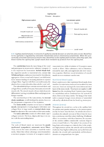
6.12 Capillary bed (schematic). A network of capillaries extends between an arteriole and a venule. Blood flow
to the capillaries is regulated by contractile metarterioles and an arteriovenous anastomosis. When the auto-
nomically controlled arteriovenous shunts close, blood flow to the capillary bed increases; when they open, less
blood reaches the capillary bed. Lymph vessels drain metabolic by-products from the capillary bed.
The endothelium forms the inner lining of the vessel vasoconstriction, while stimulation of β-receptors causes
and participates in transvascular substance transport. It vessels to dilate. Other substances, such as histamine,
is also important for haemostasis. Weibel–Palade bod- released by mast cells, and angiotensin also have vasoac-
ies, organelles specific to endothelial cells, contain von tive properties. Rhythmic neural stimulation of smooth
Willebrand factor, a substance important for thrombocyte muscle serves to maintain vascular tone.
aggregation. When a vessel wall is injured, von Willebrand
factor induces binding of circulating platelets to exposed Nutritional blood supply
collagen fibres. Together with fibrin and erythrocytes, the Blood vessels receive their nutritional blood supply from
platelets form a microthrombus that seals the vessel wall. vessels in the outer tunica media and tunica adventitia (vasa
The subendothelial layer contains scattered elastic and vasorum). Vessels are lacking in the tunica intima and inner
collagen fibres, as well as fibrocytes, histiocytes and smooth layers of the tunica media. These layers are supplied via dif-
muscle cells. The smooth muscle cells are relatively poorly fusion from the circulating blood. Nutrients pass through
differentiated, serving to synthesise fibres and phagocytose the endothelium and are distributed into the network of
foreign materials. elastic fibres in the tunica media. The lining of the vessel
The outermost layer of the tunica intima, the internal walls is renewed through mitotic division of endothelial
elastic membrane, is well developed in arteries, less so in cells and by cells derived from the blood (e.g. monocytes).
the low-pressure components of the circulation.
The tunica media comprises several layers of smooth Arteries (arteria)
muscle, interspersed to varying degrees with elastic and Throughout the arterial tree, as far as the capillary bed,
collagen fibres. In large vessels, an external elastic mem- the arteries exert passive resistance to the pressure gener-
brane lies adjacent to the outer surface of the tunica media. ated by the heart. Near the heart, the arteries also serve to
The tunica adventitia consists of a fibro-elastic (type I transform the pulsatile ejection of blood from the heart
collagen) network. This loose, displaceable layer is in con- into a continuous, even flow (elastic buffering, Windkessel
tact with the surrounding tissues. effect). In accordance with these functions, the structure
of the arteries exhibits considerable variation (Figures
Innervation 6.8 to 6.14 and Table 6.1). The walls of the arteries of the
The walls of blood vessels are innervated by poorly lung, brain and meninges are relatively thin. In the periph-
myelinated (sympathetic) autonomic nerve fibres. eral high-pressure system, the arterial wall is noticeably
These traverse the tunica adventitia to reach the tunica thickened. Near highly mobile joints, such as the knee and
media, where they release adrenergic neurotransmitters shoulder, the arterial wall is particularly well developed.
at terminal synapses. Activation of α-receptors results in Arteries are categorised into two types:
Vet Histology.indb 125 16/07/2019 14:58