
Page 221 - Veterinary Histology of Domestic Mammals and Birds, 5th Edition
P. 221
Digestive system (apparatus digestorius) 203
neck The parietal cell (exocrinocytus parietalis) is a rounded
VetBooks.ir mucous neck cells (mucocyti cervicales). Occurring indi- between, or lies peripheral to, chief cells (Figures 10.32, 10.
The neck region contains parietal cells (see below) and to pear-shaped, strongly acidophilic cell that is interposed
vidually or in groups, mucous neck cells differ in structure 33, 10.35 and 10.37). The nucleus is spherical. Parietal cells
and function to isthmus cells. Neck cells are cuboidal to occur throughout the wall of tubular glands, particularly
low columnar and typically stain only weakly. The nucleus in the middle section, and are characterised by the pres-
occupies a basal position and the accumulation of secre- ence of abundant mitochondria containing particularly
tory granules causes the apical surface of the cell to bulge numerous cristae. Tubulovesicular structures accumulate
into the lumen. beneath the plasmalemma during the inactive stage of the
The mucus produced by neck cells is acidic, of parietal cell.
relatively low viscosity and comparatively high in gly- In addition, parietal cells contain a system of intracel-
cosaminoglycans. The layer formed by the combination lular canaliculi (canaliculi intracellulares) that increases
of this mucus, and that produced by isthmus and surface the surface area across which secretion can occur. During
cells, protects the epithelium against the proteolytic and the active secretory phase, the number of microvilli lining
+
hydrolytic activity of the proteases and H ions secreted the canaliculi increases while the tubulovesicular system
by parietal cells (see below). regresses (Figure 10.35). Dissociated hydrochloric acid
+
–
(H and Cl ) is secreted into the canaliculi. Depending on
body and fundus species, parietal (and/or chief cells) also secrete intrinsic
The epithelial lining of the body and fundus of the proper factor required for absorption of vitamin B .
12
gastric glands incorporates numerous exocrine chief cells Intra-epithelial endocrine cells (endocrinocyti gas-
and parietal cells. Also present are endocrine cells of the trointestinales) are found particularly in the middle and
enteroendocrine system of the gastrointestinal tract. terminal sections of the gland. Together with endocrine
The chief cell (exocrinocytus principalis) is cuboidal cells of the pylorus and small intestine, and endocrine
or pyramidal with slightly basophilic cytoplasm (Figures cells of the pancreas, these cells belong to the gastroen-
10.32, 10.33 and 10.35). The oval nucleus is usually located teropancreatic (GEP) endocrine system. Endocrine cells
in the basal third of the cell. Typical of cells with high found in the region of the proper gastric glands include
secretory activity, chief cells are rich in organelles associ- enterochromaffin (EC) cells (serotonin), D cells (somato-
ated with protein synthesis (ribosomes, rough ER). Chief statin) and G cells (gastrin) (Figure 10.38, see also Figure
cells contain zymogen granules that, in the active secre- 2.22); their relative number varies with species.
tory phase, cause apical bulging of the cells. Chief cells
secrete pepsinogen, the precursor of pepsin.
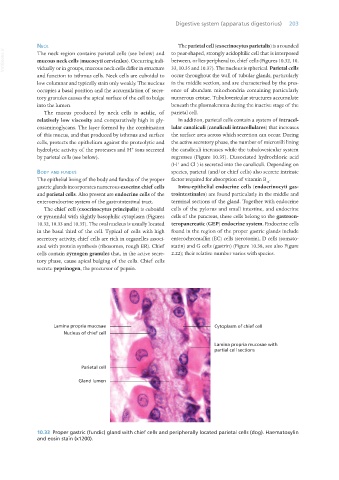
10.33 Proper gastric (fundic) gland with chief cells and peripherally located parietal cells (dog). Haematoxylin
and eosin stain (x1200).
Vet Histology.indb 203 16/07/2019 15:01