
Page 217 - Veterinary Histology of Domestic Mammals and Birds, 5th Edition
P. 217
Digestive system (apparatus digestorius) 199
The mucosa of the multi-chambered stomach of glands in the individual regions of the stomach are vari-
VetBooks.ir ruminants is non-glandular throughout the forestomach able, depending on species (Figures 10.28 to 10.38 and
(reticulum, rumen and omasum) and glandular in the Table 10.2).
The tunica mucosa is lined with simple columnar epi-
abomasum.
thelium incorporating numerous mucus-producing cells.
Glandular stomach (pars glandularis) The surface of each individual cell (epitheliocytus super-
The gastric mucosa (tunica mucosa gastrica) and under- ficialis gastricus) typically bulges towards the lumen and
lying tela submucosa are thrown into longitudinal folds is coated with a layer of highly viscous mucus that resists
(plicae gastricae). The mucosal surface is divided into the effects of gastric acids. Mucous neck cells of the gastric
areas (areae gastricae) onto which gastric pits (foveolae glands also contribute to the mucous lining. The epithelial
gastricae) open. At the base of the pits, tubular gastric cells contain abundant metabolic organelles and a central
glands extend into the lamina propria. The distribution nucleus, and are firmly held together by tight junctions
of gastric pits and the shape, size and structure of the (zonulae occludentes) and gap junctions (nexus). The
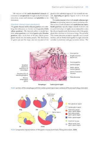
10.28 Junction of the oesophagus with the cardiac gland region (pars cardiaca) of the stomach (dog; schematic).
10.29 Comparative representation of the gastric mucosa (schematic; König and Liebich, 2009).
Vet Histology.indb 199 16/07/2019 15:01