
Page 212 - Veterinary Histology of Domestic Mammals and Birds, 5th Edition
P. 212
194 Veterinary Histology of Domestic Mammals and Birds
VetBooks.ir
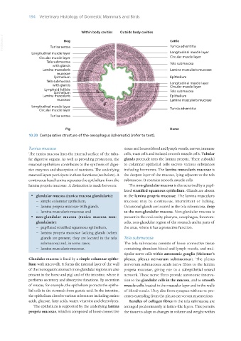
10.20 Comparative structure of the oesophagus (schematic) (refer to text).
Tunica mucosa tissue and houses blood and lymph vessels, nerves, immune
The tunica mucosa lines the internal surface of the tubu- cells, mast cells and isolated smooth muscle cells. Tubular
lar digestive organs. As well as providing protection, the glands protrude into the lamina propria. Their cuboidal
mucosal epithelium contributes to the synthesis of diges- to columnar epithelial cells secrete various substances
tive enzymes and absorption of nutrients. The underlying including hormones. The lamina muscularis mucosae is
mucosal layers participate in these functions (see below). A the deepest layer of the mucosa, lying adjacent to the tela
continuous basal lamina separates the epithelium from the submucosa. It contains smooth muscle cells.
lamina propria mucosae. A distinction is made between: The non-glandular mucosa is characterised by a papil-
lated stratified squamous epithelium. Glands are absent
· glandular mucosa (tunica mucosa glandularis): in the lamina propria mucosae. The lamina muscularis
− simple columnar epithelium, mucosae may be continuous, intermittent or lacking.
− lamina propria mucosae with glands, Occasional glands are located in the tela submucosa, deep
− lamina muscularis mucosae and to the non-glandular mucosa. Non-glandular mucosa is
· non-glandular mucosa (tunica mucosa non- present in the oral cavity, pharynx, oesophagus, forestom-
glandularis): achs, non-glandular region of the stomach and in parts of
− papillated stratified squamous epithelium, the anus, where it has a protective function.
− lamina propria mucosae lacking glands (when
glands are present, they are located in the tela Tela submucosa
submucosa) and, in some cases, The tela submucosa consists of loose connective tissue
− lamina muscularis mucosae. containing abundant blood and lymph vessels, and mul-
tipolar nerve cells within autonomic ganglia (Meissner’s
Glandular mucosa is lined by a simple columnar epithe- plexus, plexus nervorum submucosus). The plexus
lium with microvilli. It forms the internal layer of the wall nervorum submucosus sends nerve fibres to the lamina
of the monogastric stomach (non-glandular regions are also propria mucosae, giving rise to a subepithelial neural
present in the horse and pig) and of the intestine, where it network. These nerve fibres provide autonomic innerva-
performs secretory and absorptive functions. By secretion tion to the glandular cells in the mucosa, and to smooth
of mucus, for example, the epithelium protects the epithe- muscle cells located in the muscular layer and in the walls
lial cells in the stomach from gastric acid. In the intestine, of blood vessels. They also form synapses with nerve pro-
the epithelium absorbs various substances including amino cesses extending from the plexus nervorum myentericus.
acids, glucose, fatty acids, water, vitamins and electrolytes. Bundles of collagen fibres in the tela submucosa are
The epithelium is supported by the underlying lamina arranged predominantly in lattice-like layers. This permits
propria mucosae, which is composed of loose connective the tissue to adapt to changes in volume and weight within
Vet Histology.indb 194 16/07/2019 15:00