
Page 214 - Veterinary Histology of Domestic Mammals and Birds, 5th Edition
P. 214
196 Veterinary Histology of Domestic Mammals and Birds
VetBooks.ir
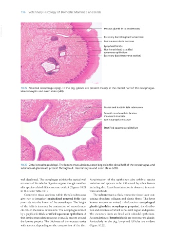
10.22 Proximal oesophagus (pig). In the pig, glands are present mainly in the cranial half of the oesophagus.
Haematoxylin and eosin stain (x40).
10.23 Distal oesophagus (dog). The lamina muscularis mucosae begins in the distal half of the oesophagus, and
submucosal glands are present throughout. Haematoxylin and eosin stain (x20).
well developed. The oesophagus exhibits the typical wall Keratinisation of the epithelium also exhibits species
structure of the tubular digestive organs, though consider- variation and appears to be influenced by other factors
able species-related differences are evident (Figures 10.21 including diet. Least keratinisation is observed in carni-
to 10.23 and Table 10.1). vores and birds.
Connective tissue cushions within the tela submucosa The submucosa is a thick connective tissue layer con-
give rise to irregular longitudinal mucosal folds that taining abundant collagen and elastic fibres. This layer
protrude into the lumen of the oesophagus. The height houses mucous or mixed, tubulo-acinar oesophageal
of the folds is increased by contraction of smooth mus- glands (glandulae oesophageae propriae), the distribu-
cle cells in the tunica muscularis. The oesophagus is lined tion and structure of which varies with region and species.
by a papillated, thick stratified squamous epithelium. A The excretory ducts are lined with cuboidal epithelium.
thin lamina muscularis mucosae is usually present around Accumulations of lymphoid cells are seen near the glands.
the lamina propria. The thickness of the mucosa varies Particularly in the pig, lymphoid follicles are evident
with species, depending on the composition of the diet. (Figure 10.22).
Vet Histology.indb 196 16/07/2019 15:00