
Page 228 - Veterinary Histology of Domestic Mammals and Birds, 5th Edition
P. 228
210 Veterinary Histology of Domestic Mammals and Birds
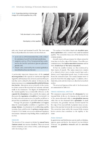
10.43 Scanning electron microscope
VetBooks.ir
image of ruminal papillae (ox; x10).
salts, urea, biuret) and vitamins B and K. The three main The surface of the folds is lined with stratified squa-
fatty acids produced in the rumen and reticulum are: mous epithelium and is covered with small keratinised
papillae. The connective tissue core of the laminae forms
· acetic acid (ca. 60% intraruminal fatty acids), utilised a lamina propria.
for synthesis of acetyl CoA for lipid metabolism, Smooth muscle cells accompany the robust connective
· propionic acid (ca. 25% of intraruminal fatty acids), tissue layer to the free edge of the laminae. These fibres are
used in the liver for synthesis of glucose (gluconeo- continuations of the lamina muscularis mucosae and the
genesis) and inner circular layer of the tunica muscularis.
· butyric acid, converted by the ruminal epithelium All but the smallest laminae are thus reinforced by
into β-hydroxybutyrate (ketone). three layers of muscle. The tunica muscularis consists
of a relatively thick inner circular muscle layer and a
A particularly important characteristic of the ruminal thinner outer longitudinal muscle layer. A tunica serosa
microorganisms is the capacity to convert non-digestible forms the outermost layer. The omasal laminae serve to
vegetable carbohydrates into ammonia and microbial pro- press the fluid from the pulpy ingesta. In addition, short-
tein that can be utilised by the animal. In this process, the chain fatty acids and electrolytes are absorbed across their
tunica mucosa acts as a semipermeable epithelial absorp- surface.
tion barrier. Absorption occurs primarily in the rumen, The structural features of the wall of the forestomach
to a lesser extent in the reticulum and omasum and mini- are summarised in Table 10.3.
mally in the abomasum. The degree of development of
the ruminal papillae has a considerable influence on the OMASO-ABOMASAL JUNCTION
capacity for absorption of fatty acids by the rumen, and The omasum is separated from the abomasum by two
thus upon the regulation and stability of ruminal pH. As mucosal folds. In the ox, the mucosa on the omasal side
the fatty acid concentration rises, the papillae proliferate to of the omaso-abomasal orifice is non-glandular mucosa
prevent the accumulation of acid in the ruminal contents. while that on the abomasal side is glandular. In small
Through the processes of proliferation and regres- ruminants, the glandular mucosa extends beyond the
sion, the ruminal papillae contribute to regulation of the free edge of the mucosal folds. Lymphatic tissue accumu-
energy balance. As absorption by proliferating papillae lates at the junction between the two mucosal epithelia.
increases, fatty acids become the primary energy source. The connective tissue within the mucosal folds contains
Control of feed intake serves as an additional regulatory barrier arteries that contribute to the rigidity of the
mechanism: ruminants stop feeding when ruminal pH is tissue.
too low.
Avian stomach
OMASUM In granivorous and herbivorous species such as chickens,
The interior of the omasum is divided by omasal leaves pigeons, geese and ducks, the stomach has two distinct
(laminae omasi) into narrow chambers. There are several divisions, the glandular stomach and the muscular
orders of laminae (one to four or five), each subsequent stomach.
order decreasing in size.
Vet Histology.indb 210 16/07/2019 15:01