
Page 232 - Veterinary Histology of Domestic Mammals and Birds, 5th Edition
P. 232
214 Veterinary Histology of Domestic Mammals and Birds
nucleases and phosphatases). In addition, bile produced by pinocytosis and converted into triglycerides in the smooth
VetBooks.ir the liver and, in some cases, enzymes present in foodstuffs, endoplasmic reticulum. These are furnished with a lipo-
contribute to the digestive process.
protein coat in the Golgi apparatus and transported as
Food components are hydrolysed to form low- chylomicrons through the intracellular compartment to
molecular weight substrates. The associated metabolic pro- the lymphatic capillaries.
cesses are regulated by neural and hormonal mechanisms. As well as secreting digestive enzymes and absorb-
In animals with simple stomachs, breakdown of pro- ing the products of digestion, the epithelial cells – to an
teins into amino acids is completed in the small intestine. even greater extent than in the stomach – synthesise and
Polypeptides produced by the action of pepsin on proteins secrete mucus for cytoprotection of the intestinal surface.
in the stomach are hydrolysed in the small intestine by In addition, tissue hormones are produced by the mucosa,
trypsin and chymotrypsin, giving rise to oligopeptides, and primarily by the epithelial cells of the intestinal glands.
by peptidases to produce L-amino acids. Polysaccharides The mucosa also absorbs water, electrolytes and vitamins.
are broken down by pancreatic α-amylase and intestinal Furthermore, in terms of surface area, the enteral mucosa
1,6-glucosidase into monosaccharides (maltose, isomalt- constitutes the largest lymphatic organ of the body and
ose, glucose). Following emulsification in the stomach, actively participates in immune responses.
triglycerides are cleaved in the small intestine by pan- The small intestine exhibits the typical layered muscu-
creatic lipase to form β-monoglycerides and fatty acids. lomembranous structure of the tubular digestive organs.
Digestion of fats is further aided by bile salts and cho- Apart from certain segment-specific differences, the mor-
lesterol. Additional pancreatic enzymes (nucleases and phology of the small intestine is similar, and the following
phosphatases) catalyse the conversion of nucleic acids into description applies to the duodenum, jejunum and ileum.
nuclear bases and pentoses. A summary of the structural features of the segments
These processes rely on continuous mixing of chyme of the intestine is provided in Table 10.5.
with hydrolytic secretions that function optimally in a
neutral to slightly alkaline environment. End products Tunica mucosa
available for absorption are taken up at the luminal sur- The various functions of the small intestine depend
face of the mucosal epithelium and pass through the basal largely on structural specialisations that greatly increase
portion of the cell to reach the blood or lymph system. its internal surface area. These include transversely ori-
Absorption is an active process that utilises concen- ented macroscopically visible folds (plicae circulares) that
tration gradients and/or the membrane potential of the gradually decrease in height in the caudal segments of the
plasmalemma. intestine. In ruminants, the folds are permanent. The core
Short-chain fatty acids formed by the hydrolysis of of the folds is composed of loose connective tissue in the
fats pass through the epithelium into the blood capillaries tela submucosa that protrudes into the intestinal lumen.
and then directly to the liver. Long-chain fatty acids and In addition, the entire small intestinal mucosa is covered
monoglycerides are taken up into the epithelial cells by with intestinal villi (villi intestinales), finger-like evagina-
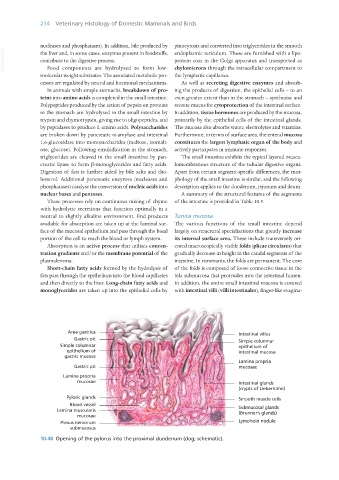
10.48 Opening of the pylorus into the proximal duodenum (dog; schematic).
Vet Histology.indb 214 16/07/2019 15:01