
Page 235 - Veterinary Histology of Domestic Mammals and Birds, 5th Edition
P. 235
Digestive system (apparatus digestorius) 217
In addition to the projections from its surface, the mucosa protection of the intestinal surface by mucus is typically lack-
VetBooks.ir is characterised by invaginations of the lamina propria in ing at this time, the protracted cellular lifespan can increase
the form of straight, non-branching tubular intestinal vulnerability to gastrointestinal infection (e.g. diarrhoea)
glands (glandulae intestinales, crypts of Lieberkühn) in newborn animals. As the intestinal glands also undergo
(Figures 10.50 and 10.54 to 10.57). Undifferentiated epi- postnatal development, the capacity for rapid replacement
thelial cells (epitheliocyti nondifferentiati) at the base of shed epithelial cells is not yet fully developed.
of the glands undergo constant mitotic division. Daughter The intestinal villi and the walls of the intestinal glands
cells migrate towards the lumen to the tip of the intestinal are lined by a simple columnar epithelium incorporating
villi where they replace continuously desquamating cells, several cell types that vary in structure and function:
thus serving to regenerate the absorptive surface.
The lifespan of an intestinal epithelial cell is generally · enterocytes (epitheliocyti columnares villi),
10–14 days in neonates and 2–5 days in adults. The relatively · goblet cells (epitheliocyti caliciformes),
long lifespan in juveniles facilitates intra-epithelial mul- · endocrine cells (endocrinocyti gastrointestinales) and
tiplication of microbial pathogens within the mucosa. As · Paneth cells (exocrinocyti cum granulis acidophilis).
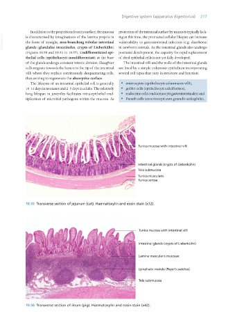
10.55 Transverse section of jejunum (cat). Haematoxylin and eosin stain (x12).
10.56 Transverse section of ileum (pig). Haematoxylin and eosin stain (x42).
Vet Histology.indb 217 16/07/2019 15:01