Page 236 - Veterinary Histology of Domestic Mammals and Birds, 5th Edition
P. 236
218  Veterinary Histology of Domestic Mammals and Birds


       VetBooks.ir


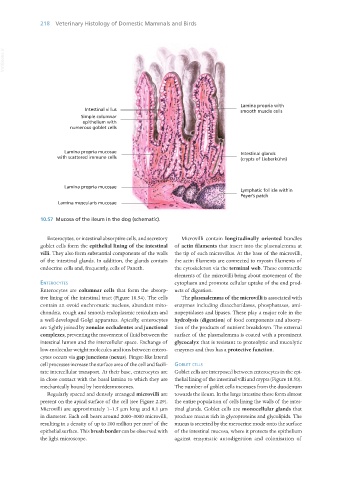
                   10.57  Mucosa of the ileum in the dog (schematic).


                      Enterocytes, or intestinal absorptive cells, and secretory   Microvilli contain  longitudinally oriented  bundles
                   goblet cells form the epithelial lining of the intestinal  of actin filaments that insert into the plasmalemma at
                   villi. They also form substantial components of the walls  the tip of each microvillus. At the base of the microvilli,
                   of the intestinal glands. In addition, the glands contain  the actin filaments are connected to myosin filaments of
                   endocrine cells and, frequently, cells of Paneth.  the cytoskeleton via the terminal web. These contractile
                                                                  elements of the microvilli bring about movement of the
                   enteRocytes                                    cytoplasm and promote cellular uptake of the end prod-
                   Enterocytes are columnar cells that form the absorp-  ucts of digestion.
                   tive lining of the intestinal tract (Figure 10.54). The cells   The plasmalemma of the microvilli is associated with
                   contain an ovoid euchromatic nucleus, abundant mito-  enzymes including disaccharidases, phosphatases, ami-
                   chondria, rough and smooth endoplasmic reticulum and  nopeptidases and lipases. These play a major role in the
                   a well-developed Golgi apparatus. Apically, enterocytes  hydrolysis (digestion) of food components and absorp-
                   are tightly joined by zonulae occludentes and junctional  tion of the products of nutrient breakdown. The external
                   complexes, preventing the movement of fluid between the  surface of the plasmalemma is coated with a prominent
                   intestinal lumen and the intercellular space. Exchange of  glycocalyx that is resistant to proteolytic and mucolytic
                   low-molecular weight molecules and ions between entero-  enzymes and thus has a protective function.
                   cytes occurs via gap junctions (nexus). Finger-like lateral
                   cell processes increase the surface area of the cell and facili-  goblet cells
                   tate intercellular transport. At their base, enterocytes are  Goblet cells are interposed between enterocytes in the epi-
                   in close contact with the basal lamina to which they are  thelial lining of the intestinal villi and crypts (Figure 10.50).
                   mechanically bound by hemidesmosomes.          The number of goblet cells increases from the duodenum
                      Regularly spaced and densely arranged microvilli are  towards the ileum. In the large intestine these form almost
                   present on the apical surface of the cell (see Figure 2.29).  the entire population of cells lining the walls of the intes-
                   Microvilli are approximately 1–1.5 μm long and 0.1 μm  tinal glands. Goblet cells are monocellular glands that
                   in diameter. Each cell bears around 2000–3000 microvilli,  produce mucus rich in glycoproteins and glycolipids. The
                   resulting in a density of up to 200 million per mm  of the  mucus is secreted by the merocrine mode onto the surface
                                                          2
                   epithelial surface. This brush border can be observed with  of the intestinal mucosa, where it protects the epithelium
                   the light microscope.                          against enzymatic autodigestion and colonisation of


       Vet Histology.indb   218                                                                                  16/07/2019   15:01

