
Page 350 - Veterinary Histology of Domestic Mammals and Birds, 5th Edition
P. 350
332 Veterinary Histology of Domestic Mammals and Birds
tubular glands (Figure 15.12). The epithelial lining of the pyknotic and the organelles degenerate. As production of
VetBooks.ir tubular secretory portion is cuboidal to columnar, depend- new cells continues, older cells are forced into the lumen of
ing on secretory activity. The ducts open independently of the alveolar gland where they break down. The cell debris
hair follicles directly onto the surface of the skin. Release and secretory product together form sebum. Sebaceous
of secretory products into the tubular lumen is facilitated glands may be simple or compound. They also occur inde-
by myoepithelial cells that lie on the deep aspect of the pendently of hair follicles, e.g. in the eyelid (Meibomian
secretory units. These are modified epithelial cells that glands), in the prepuce and penis, and in the external ear
have acquired the ability to contract. canal.
Sweat glands have several functions. They contribute The secretory product of sebaceous glands spreads over
to thermoregulation through evaporation of sweat and the surface of the skin, covering the entire epidermis with
provide a means of eliminating products of metabolism. a thin layer of lipid. It serves as a waterproofing agent,
Their secretions mix with those of sebaceous glands form- has an antimicrobial function and lubricates the stratum
ing a film on the surface of the skin. In domestic mammals corneum of the epidermis and the hair.
this layer is only weakly acidic, becoming alkaline with Numerous modified sweat and sebaceous glands are
increased output from the apocrine sweat glands. In con- found in domestic mammals. The structure of the glands
trast to the hairless skin of humans, this layer does not of the planum nasolabiale of ruminants, glands of the anal
provide protection in the form of an acidic environment. sacs of carnivores and the circumanal glands of dogs is
The antimicrobial effect of the layer coating the skin sur- described in Chapter 10, ‘Digestive system’.
face is probably related to free fatty acids, formed during
the decomposition of epidermal surface lipids. Pigmentation
Even in densely haired regions, the presence of pigments
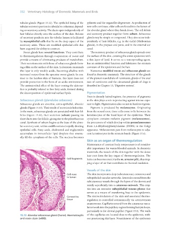
Sebaceous glands (glandulae sebaceae) in the skin plays a role in protecting against intense expo-
Sebaceous glands are exocrine, extra-epithelial, alveolar sure to light. Pigmentation also occurs in hairless regions.
glands (Figure 15.13). Their mode of secretion is holocrine. Pigment is produced by melanocytes. Originating
In the main, sebaceous glands are associated with hair fol- from the neural crest, these cells come to lie between the
licles (Figure 15.10), their secretion (sebum) passing via keratinocytes of the basal layer of the epidermis. Their
short ducts into the follicle, giving rise to the pilosebaceous cytoplasm contains melanin pigment (melanosomes),
canal. Synthesis of sebum begins at the base of the alveo- the precursors of which develop within premelanosomes
lar secretory unit, within undifferentiated rapidly dividing from 3,4-dihydroxyphenylalanine (DOPA) in the Golgi
epithelial cells. Fatty acids, cholesterol and triglycerides apparatus. Melanosomes pass from melanocytes to adja-
accumulate in intracellular lipid droplets that eventu- cent keratinocytes in the stratum basale (Figure 15.6).
ally fill the cytoplasm of the cells. The nucleus becomes
Skin as an organ of thermoregulation
Maintenance of constant body temperature is of consider-
able importance for warm-blooded animals. In domestic
mammals, the vessels of the skin together with the dense
hair coat form the key organ of thermoregulation. The
Cell fragments hairs can become erect (via the m. arrector pili), thus trap-
ping a layer of air that contributes to thermal insulation.
Vessels of the skin
Cell mass The skin incorporates deep (subcutaneous), cutaneous and
in lumen subepithelial vascular networks. Arterioles extend from the
subcutaneous vessels through the layers of the dermis and
ramify superficially into a cutaneous network. This emp-
ties into an extensive subepithelial venous plexus that
serves as a means of transferring heat to the epidermis.
Connective The microcirculation of the skin and associated thermo-
tissue regulation is controlled autonomically via arteriovenous
anastomoses. Capillaries extend from the cutaneous vascu-
lar network into the papillary region forming hairpin loops,
particularly in the dermal papillae (Figure 15.8). The walls
15.13 Alveolar sebaceous gland (horse). Haematoxylin of the capillaries are located close to the epidermis, with-
and eosin stain (x400). out penetrating this layer. Nourishment of the epidermis
Vet Histology.indb 332 16/07/2019 15:06