
Page 346 - Veterinary Histology of Domestic Mammals and Birds, 5th Edition
P. 346
328 Veterinary Histology of Domestic Mammals and Birds
vation are well developed (see below). It is particularly in
· melanocytes (pigment cells), protective function,
VetBooks.ir · Langerhans cells (macrophages), immune function this layer that epidermal appendages such as hairs, and
sebaceous and sweat glands are embedded (see below).
and
The papillary layer is composed of loose connective
· Merkel cells, sensory (tactile) function.
tissue, comprising a lattice of collagen fibres interwoven
with elastic fibres, the latter condensing to form a strong
Dermis (corium) subepithelial layer. (Figure 15.7). Type III collagen fibres
The dermis is the connective tissue layer that underlies interweave with the basal lamina, forming a functional
the epidermis (Figures 15.2 and 15.5 to 15.10). Based upon connection with the network of tonofibrils within the epi-
variations in density and the arrangement of its scaffold thelial keratinocytes. This close association between the
of collagen and elastic fibres, the dermis is divided into papillary layer and the basal cells of the epidermis imparts
two layers: considerable mechanical strength to the outer layers of
the skin.
· stratum papillare (papillary layer) with: Hairpin-shaped capillary loops sprout into the dermal
− dermal papillae, papillae, supplying blood to the epidermis (Figure 15.8).
− connective tissue fibres, hair, sebaceous and Interdigitation of the papillary layer with the epidermis
sweat glands, facilitates diffusion of nutrients into the epidermis in
− microvasculature (capillary loops), general, and the regenerative capacity of the stratum ger-
− autonomic innervation and minativum in particular.
· stratum reticulare (reticular layer) containing a lat- The loose connective tissue of the papillary layer is
tice of connective tissue fibres. interspersed with lymphocytes, plasma cells, macrophages
and mast cells. The presence of considerable numbers of
these cells is indicative of adaptive and innate immune
Papillary layer (stratum papillare) responses that occur continuously within the skin.
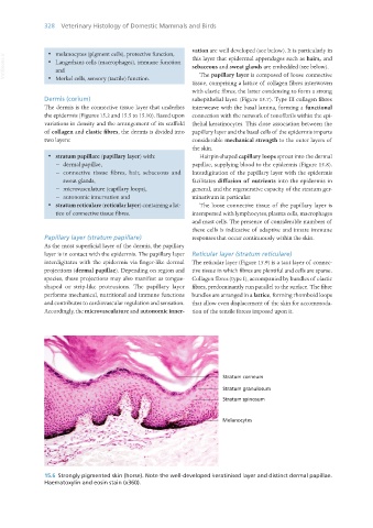
As the most superficial layer of the dermis, the papillary
layer is in contact with the epidermis. The papillary layer Reticular layer (stratum reticulare)
interdigitates with the epidermis via finger-like dermal The reticular layer (Figure 15.9) is a taut layer of connec-
projections (dermal papillae). Depending on region and tive tissue in which fibres are plentiful and cells are sparse.
species, these projections may also manifest as tongue- Collagen fibres (type I), accompanied by bundles of elastic
shaped or strip-like protrusions. The papillary layer fibres, predominantly run parallel to the surface. The fibre
performs mechanical, nutritional and immune functions bundles are arranged in a lattice, forming rhomboid loops
and contributes to cardiovascular regulation and sensation. that allow even displacement of the skin for accommoda-
Accordingly, the microvasculature and autonomic inner- tion of the tensile forces imposed upon it.
15.6 Strongly pigmented skin (horse). Note the well-developed keratinised layer and distinct dermal papillae.
Haematoxylin and eosin stain (x360).
Vet Histology.indb 328 16/07/2019 15:06