
Page 344 - Veterinary Histology of Domestic Mammals and Birds, 5th Edition
P. 344
326 Veterinary Histology of Domestic Mammals and Birds
the identification of epithelial tissue and its stage of dif-
VetBooks.ir ferentiation in the diagnosis of neoplasia. Cytokeratins
are assembled in a specific manner, depending upon the
functional demands on the epithelium. Accordingly, epi-
dermal regions and structures such as hairy skin, digital
pads and the claws each exhibit a characteristic cytokeratin
pattern.
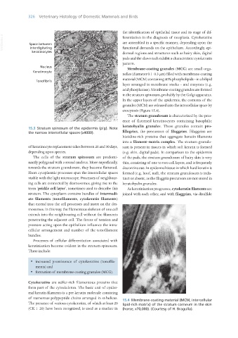
Membrane-coating granules (MCG) are small orga-
nelles (diameter 0.1–0.3 μm) filled with membrane-coating
material (MCM) containing 40% phospholipids – in a bilipid
layer arranged in membrane stacks – and enzymes (e.g.
acid phosphatase). Membrane-coating granules are formed
in the stratum spinosum, probably by the Golgi apparatus.
In the upper layers of the epidermis, the contents of the
granules (MCM) are released into the intercellular space by
exocytosis (Figure 15.4).
The stratum granulosum is characterised by the pres-
ence of flattened keratinocytes containing basophilic
keratohyalin granules. These granules contain pro-
15.3 Stratum spinosum of the epidermis (pig). Note
the narrow intercellular spaces (x4000). fillagrins, the precursors of filaggrins. Filaggrins are
histidine-rich proteins that aggregate keratin filaments
into a filament–matrix complex. The stratum granulo-
of keratinocyte replacement takes between 20 and 30 days, sum is present in tissues in which soft keratin is formed
depending upon species. (e.g. skin, digital pads). In comparison to the epidermis
The cells of the stratum spinosum are predomi- of the pads, the stratum granulosum of hairy skin is very
nantly polygonal with a round nucleus. More superficially, thin, consisting of one to two cell layers, and is frequently
towards the stratum granulosum, they become flattened. discontinuous. In epidermal tissue in which hard keratin is
Short cytoplasmic processes span the intercellular spaces formed (e.g. hoof, nail), the stratum granulosum is indis-
visible with the light microscope. Processes of neighbour- tinct or absent, as the filaggrin precursors are not stored in
ing cells are connected by desmosomes, giving rise to the keratohyalin granules.
term ‘prickle cell layer’, sometimes used to describe this As keratinisation progresses, cytokeratin filaments are
stratum. The cytoplasm contains bundles of intermedi- joined with each other, and with filaggrins, via disulfide
ate filaments (tonofilaments, cytokeratin filaments)
that extend into the cell processes and insert on the des-
mosomes. In this way, the filamentous skeleton of one cell
extends into the neighbouring cell without the filaments
penetrating the adjacent cell. The forces of tension and
pressure acting upon the epithelium influence the intra-
cellular arrangement and number of the tonofilament
bundles.
Processes of cellular differentiation associated with
keratinisation become evident in the stratum spinosum.
These include:
· increased prominence of cytokeratins (tonofila-
ments) and
· formation of membrane-coating granules (MCG).
Cytokeratins are sulfur-rich filamentous proteins that
form part of the cytoskeleton. The basic unit of epider-
mal keratin filaments is a pre-keratin molecule consisting
of numerous polypeptide chains arranged in α-helices. 15.4 Membrane-coating material (MCM, intercellular
The presence of various cytokeratins, of which at least 20 lipid-rich matrix) of the stratum corneum in the skin
(CK 1–20) have been recognised, is used as a marker in (horse; x70,000). (Courtesy of H. Bragulla).
Vet Histology.indb 326 16/07/2019 15:06