
Page 343 - Veterinary Histology of Domestic Mammals and Birds, 5th Edition
P. 343
Common integument (integumentum commune) 325
In a stricter sense, the epidermis and dermis may be con- surface, the cells undergo multiple stages of differen-
VetBooks.ir sidered to constitute the skin, which is underlaid by the tiation associated with the process of keratinisation,
subcutis. The layers of the skin are further subdivided as culminating in the formation of dead, keratinised
cells.
follows:
The various layers each perform important functions
· epidermis: in the continuous process of formation, keratinisation and
− stratum corneum (keratinised layer), ultimately removal (desquamation) of keratinocytes. The
− stratum lucidum (eleidin layer), stratum basale serves as a germinal layer in which new
− stratum granulosum (formation of keratohyalin keratinocytes are constantly produced. For this reason, it is
granules), also referred to as the stratum germinativum. The newly
− stratum spinosum (keratinocytes), formed cells move into the stratum spinosum (Figure
− stratum basale (keratinocytes), 15.3), where changes associated with keratinisation begin
· dermis (corium): to occur. Keratinisation takes place in the transition from
− stratum papillare and the stratum granulosum to (in some cases) the stratum
− stratum reticulare. lucidum and to the stratum corneum. In the stratum
corneum, the keratinocytes are packed together to form
a dense cornified band that serves as the protective epider-
Skin as a protective organ mal barrier of the skin.
The stratum basale consists of a single layer of
Epidermis cuboidal to columnar cells that rests on a basal lamina.
As the most superficial layer of the skin, the epidermis pro- The cells are connected to the basal lamina by hemides-
tects the organism against damage caused by mechanical, mosomes, firmly anchoring the epidermis to the dermis.
thermal, chemical and biological influences (Figures 15.2 This mechanical connection is reinforced by tight inter-
to 15.8). It consists of stratified squamous epithelium digitations between dermal papillae and complementary
that exhibits regional variation in cornification, as deter- epidermal invaginations (epidermal pegs). Formation
mined by prevailing mechanical forces (see also Chapter 2, of new keratinocytes in the stratum basale occurs by
‘Epithelial tissue’). Approximately 85% of the cells of the mitosis. The rate of cell division is influenced by the rate
epidermis are keratinocytes that move from the base to of desquamation and by mechanical wear and tear of the
the surface of the epithelium. As they progress to the cornified cells at the surface of the epithelium. A full cycle
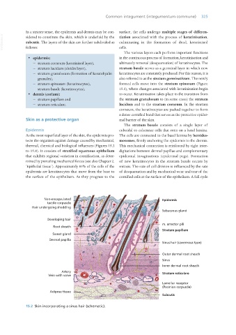
Non-encapsulated Epidermis
tactile corpuscle
Hair undergoing shedding
Sebaceous gland
Developing hair
m. arrector pili
Root sheath
Stratum papillare
Sweat gland
Dermal papilla
Sinus hair (cavernous type)
Outer dermal root sheath
Sinus
Inner dermal root sheath
Artery Stratum reticulare
Vein with valve
Lamellar receptor
(Pacinian corpuscle)
Adipose tissue
Subcutis
15.2 Skin incorporating a sinus hair (schematic).
Vet Histology.indb 325 16/07/2019 15:06