Page 345 - Veterinary Histology of Domestic Mammals and Birds, 5th Edition
P. 345
Common integument (integumentum commune)   327


       VetBooks.ir


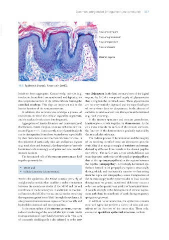
                  15.5  Epidermis (horse). Azan stain (x400).

                  bonds to form aggregates. Concurrently, proteins (e.g.  tum disjunctum. In the hard coronary horn of the digital
                  involucrin, keratolinin) are synthesised and deposited on  organs, the MCM is composed largely of glycoproteins
                  the cytoplasmic surface of the cell membrane forming the  that strengthen the cornified tissue. These glycoproteins
                  cornified envelope. This plays an important role in the  are not enzymatically degraded and the superficial layer
                  barrier function of the stratum corneum.       of horny tissue does not desquamate. In the absence of
                     In addition, the keratinocytes undergo a process of  sufficient natural wear and tear, this layer must be trimmed
                  necrobiosis, in which the cellular organelles degenerate,  (e.g. hoof trimming).
                  and the nucleus breaks down into fragments.       In the stratum spinosum and stratum granulosum,
                     Aggregation of keratin filaments and condensation of  keratinocytes are held together by desmosomes. As the
                  the filament–matrix complex continues in the stratum cor-  cells move towards the surface of the stratum corneum,
                  neum (Figure 15.5). Consequently, newly keratinised cells  the function of the desmosomes is gradually replaced by
                  can be distinguished from those located more superficially  the intercellular substance.
                  by their histochemical and mechanical characteristics. In   The ordered process of keratinisation and the integrity
                  the epidermis of particularly thick skin and hairless regions  of the resulting cornified tissue are dependent upon the
                  (e.g. nasal plane and foot pads), the deeper layer of recently  availability of an adequate supply of nutrients and energy,
                  keratinised cells is strongly acidophilic and is termed the   derived by diffusion from vessels in the dermal papillae
                  stratum lucidum.                               (see below). The surface area across which diffusion can
                     The keratinised cells of the stratum corneum are held  occur is greater on the sides of the papillae (peripapillary)
                  together primarily by:                         than at the tips (suprapapillary) or the regions between
                                                                 the papillae (interpapillary). Accordingly, keratinised epi-
                   ·  MCM and                                    thelium formed in the peripapillary region is structurally
                   ·  cellular junctions (desmosomes).           distinguishable and mechanically superior to that arising
                                                                 from the supra- and interpapillary zones. Compromise of
                  Within the epidermis, the MCM  consists primarily of  the nutrient supply to the epidermis due to local vascular
                  acylglucosylceramides that establish a stable connection  derangement or general nutritional deficiency causes a
                  between the membrane stacks of the MCM and the cell  reduction in the quantity and quality of keratinised tissue.
                  membrane of the keratinocytes. In addition to its mechani-  A notable example is the development of circular depres-
                  cal function, the MCM serves as a crucial barrier protecting  sions in the hard keratin (horn) of cattle during pregnancy
                  the organism against loss of fluid through the epidermis. It  (pregnancy grooves).
                  also prevents transcutaneous ingress of water-soluble and   In addition to keratinocytes, the epidermis contains
                  hydrophilic chemicals and microorganisms.      other cell types that perform a variety of roles and con-
                     At the outer surface of the stratum corneum, enzyme-  tribute to the function of the outer skin. These cells,
                  induced weakening of the intercellular lipid matrix results  considered specialised epidermal structures, include:
                  in desquamation of superficial keratinised cells. This layer
                  of constantly shedding cells is also referred to as the stra-


       Vet Histology.indb   327                                                                                  16/07/2019   15:06

