
Page 47 - Veterinary Histology of Domestic Mammals and Birds, 5th Edition
P. 47
The cell (cellula) 29
VetBooks.ir Leptotene S phase
S phase
Zygotene
Pachytene Prophase
Diplotene
Diakinsis
Metaphase Metaphase
Anaphase Anaphase
Telophase Telophase
Meiosis I
Meiosis II
G phase G phase
0 1
Meiosis Mitosis
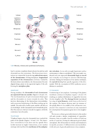
1.39 Meiosis, mitosis and cytokinesis (schematic).
bind to protein complexes (kinetochores) located on each mic reticulum. An ion-rich, strongly hypertonic nuclear
chromatid near the centromere. The kinetochore micro- environment is thus re-established. The previously con-
tubules are responsible for producing ordered movement densed and now separated chromatids begin to uncoil
of the chromosomes. During metaphase, longitudinal and eventually can no longer be detected with histologi-
separation of the chromosomes continues. Guided by cal stains. During the formation of the nuclear envelopes,
the kinetochore microtubules, the chromosomes become the nucleoli develop from specific segments of the chro-
aligned along a plane in the equatorial region of the cell, matids. With the resumption of RNA synthesis, nuclear
forming the metaphase plate. division is concluded.
ANAPHASE CYTOKINESIS
During anaphase, the chromatids of each chromosome Commencing in late anaphase, furrowing of the plasma
are separated from one another (Figures 1.38 and 1.39). membrane continues in telophase (Figures 1.38 and 1.39)
The two kinetochores of each chromosome break apart resulting in random distribution of organelles between
and the chromatids are drawn towards the poles. This the two daughter cells. The cleavage furrow is produced
involves shortening of the kinetochore microtubules, by a ring of actin filaments, which forms at the level of
with concurrent lengthening of the fibres making up the the equator. The furrow deepens until the remains of
spindle. The poles move further apart, and the cell takes on the spindle fibres in the middle of the cell become com-
an ovoid shape. As the chromatids move towards the poles, pressed. This compacted central area is only maintained
their free ends are oriented towards the equator, while the for a short period. Its separation gives rise to two daughter
centromere faces the poles. cells.
The two new cells are genetically identical to the mother
TELOPHASE cell and contain a similar complement of organelles.
Telophase begins when the chromatids have reached the However, these are smaller than the mother cell and may
poles of the spindle (Figures 1.38 and 1.39). The kineto- not be of equal size. During the subsequent growth phase
chore microtubules break down and the spindle fibres (interphase), the cell increases in volume and commences
further increase in length. Concurrently, a new nuclear its specific processes of differentiation and metabolism.
envelope is formed from the membranes of the endoplas- A full cell cycle of growth and division is thus complete.
Vet Histology.indb 29 16/07/2019 14:53