
Page 43 - Veterinary Histology of Domestic Mammals and Birds, 5th Edition
P. 43
The cell (cellula) 25
nuclear matrix is the fluid component of the interior of During mitosis these undergo additional condensation to
VetBooks.ir the nucleus (consisting of soluble RNA, ions, glycoproteins form a discrete chromosome (Figure 1.35).
The term chromosome refers to the DNA molecule
and metabolites) in which the chromatin and nucleoli are
and its associated protein scaffold. Depending on the
embedded.
stage of the cell cycle, the structural organisation of the
Chromatin (chromatinum) chromosome varies, ranging from chromatin fibres to the
The term chromatin encompasses all of the compo- condensed form that manifests during mitosis. The genetic
nents of the chromosomal DNA within the nucleus. Also information contained within the chromosome is carried
referred to as DNA–protein (nucleoprotein) complexes, by the DNA molecule.
these components consist of approximately one-third During interphase, chromatin can be subdivided based
DNA and two-thirds basic and acidic proteins (histones on morphological criteria (independently of association
and non-histones), phospholipids, ions (Ca ), glycopro- with a particular chromosome) into euchromatin and
2+
teins and small quantities of RNA. heterochromatin.
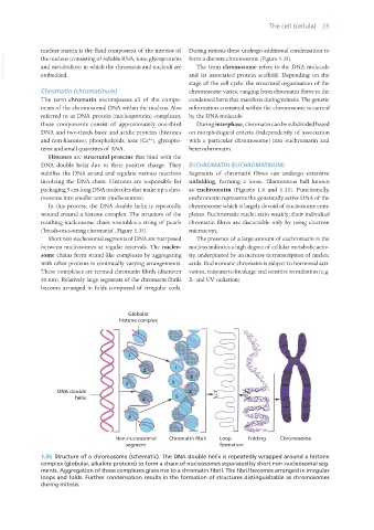
Histones are structural proteins that bind with the
DNA double helix due to their positive charge. They EUCHROMATIN (EUCHROMATINUM)
stabilise the DNA strand and regulate various reactions Segments of chromatin fibres can undergo extensive
involving the DNA chain. Histones are responsible for unfolding, forming a loose, filamentous ball known
packaging 5 cm-long DNA molecules that make up a chro- as euchromatin (Figures 1.6 and 1.33). Functionally,
mosome into smaller units (nucleosomes). euchromatin represents the genetically active DNA of the
In this process, the DNA double helix is repeatedly chromosome which is largely devoid of nucleosome com-
wound around a histone complex. The structure of the plexes. Euchromatic nuclei stain weakly; their individual
resulting nucleosome chain resembles a string of pearls chromatin fibres are discernible only by using electron
(‘beads-on-a-string chromatin’, Figure 1.35). microscopy.
Short non-nucleosomal segments of DNA are interposed The presence of a large amount of euchromatin in the
between nucleosomes at regular intervals. The nucleo- nucleus indicates a high degree of cellular metabolic activ-
some chains form strand-like complexes by aggregating ity, underpinned by an increase in transcription of nucleic
with other proteins in continually varying arrangements. acids. Euchromatic chromatin is subject to hormonal acti-
These complexes are termed chromatin fibrils (diameter vation, resistant to breakage and sensitive to radiation (e.g.
30 nm). Relatively large segments of the chromatin fibrils X- and UV radiation).
become arranged in folds composed of irregular coils.
1.35 Structure of a chromosome (schematic). The DNA double helix is repeatedly wrapped around a histone
complex (globular, alkaline proteins) to form a chain of nucleosomes separated by short non-nucleosomal seg-
ments. Aggregation of these complexes gives rise to a chromatin fibril. The fibril becomes arranged in irregular
loops and folds. Further condensation results in the formation of structures distinguishable as chromosomes
during mitosis.
Vet Histology.indb 25 16/07/2019 14:53